Thoracic Aorta - Chronic dilatation
How to measure
Lauran Stöger¹ and Robin Smithuis²
¹Utrecht University Medical Center and ²Alrijne hospital in Leiden, the Netherlands
Publicationdate
Precise measurements of the diameter of the thoracic aorta are important for detection and risk stratification of aneurysms, while accurate aortic annulus sizing is essential in Transcatheter aortic valve implantation or replacement (TAVI/TAVR).
When following up a patient with a dilated thoracic aorta over time, it is essential that serial measurements be obtained using a consistent measurement methodology.
In this article we will discuss how to measure the thoracic aorta with special attention to the aortic root.
Introduction
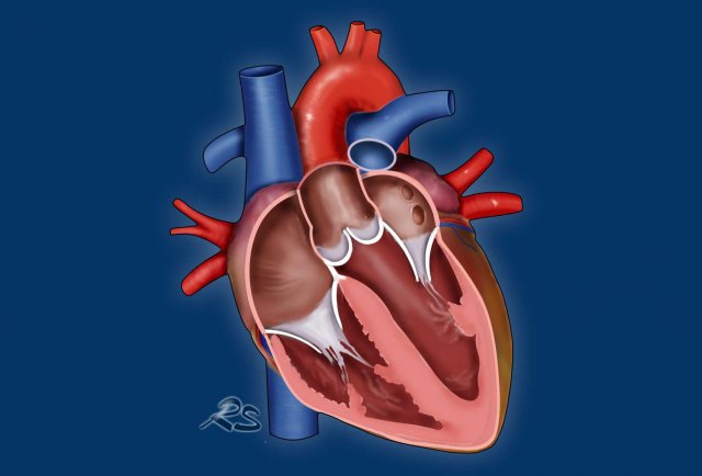
Several professional societies have issued guidelines on aortic care, detailing how the aorta should be measured. However, these guidelines differ in measurement methodology, as well as in the number and location of the measurements required.
Therefore, radiologists, cardiologists, and cardiothoracic surgeons within each institution should agree on a standardized protocol specifying how the thoracic aorta is to be measured, thereby ensuring consistency, reproducibility, and accurate longitudinal follow-up.
Dilatation of the thoracic aorta is often incidentally detected during transthoracic echocardiography (TTE) or chest CT performed for unrelated indications.
TTE measurements
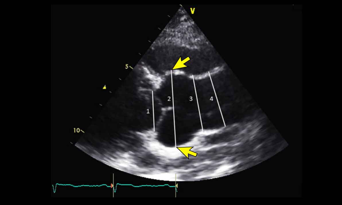
TTE measurements of the thoracic aorta are obtained using the leading edge-to-leading edge technique in end-diastole on a parasternal long-axis view.
This refers to the measurement of the diameter from the interface between the right ventricular outflow tract and the anterior aortic wall to the interface of the aortic lumen with the posterior aortic wall, as indicated by the arrows.
On TTE it is not possible to reliably measure the thickness of the posterior aortic wall.
For initial diagnosis of aortic root or proximal thoracic aorta dilatation measurements should be confirmed using cardiac CTA (CCTA).
In certain cases, magnetic resonance angiography (MRA) may be preferable to CTA, particularly in young patients or those requiring long-term follow-up.
Routine follow-up is typically performed using CTA; however, TTE may suffice as long as there is no significant discrepancy with CTA measurements and the degree of dilatation is moderate.
Matter of Millimeters
Appropriate management of thoracic aortic disease is a matter of millimeters.
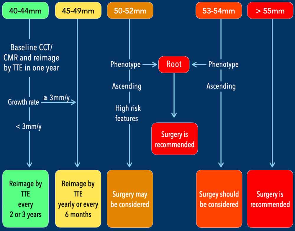
To demonstrate the importance of precise measurements, see the table on the left, representing one of the many follow-up protocols that exist for dilatation of the thoracic aorta (ESC 2024 guideline).
Do not study this table, it is only to show you that a difference of just 2 mm can reclassify a patient into a neighbouring risk category, thereby altering their follow-up protocol or surgical indication.
Normal diameters
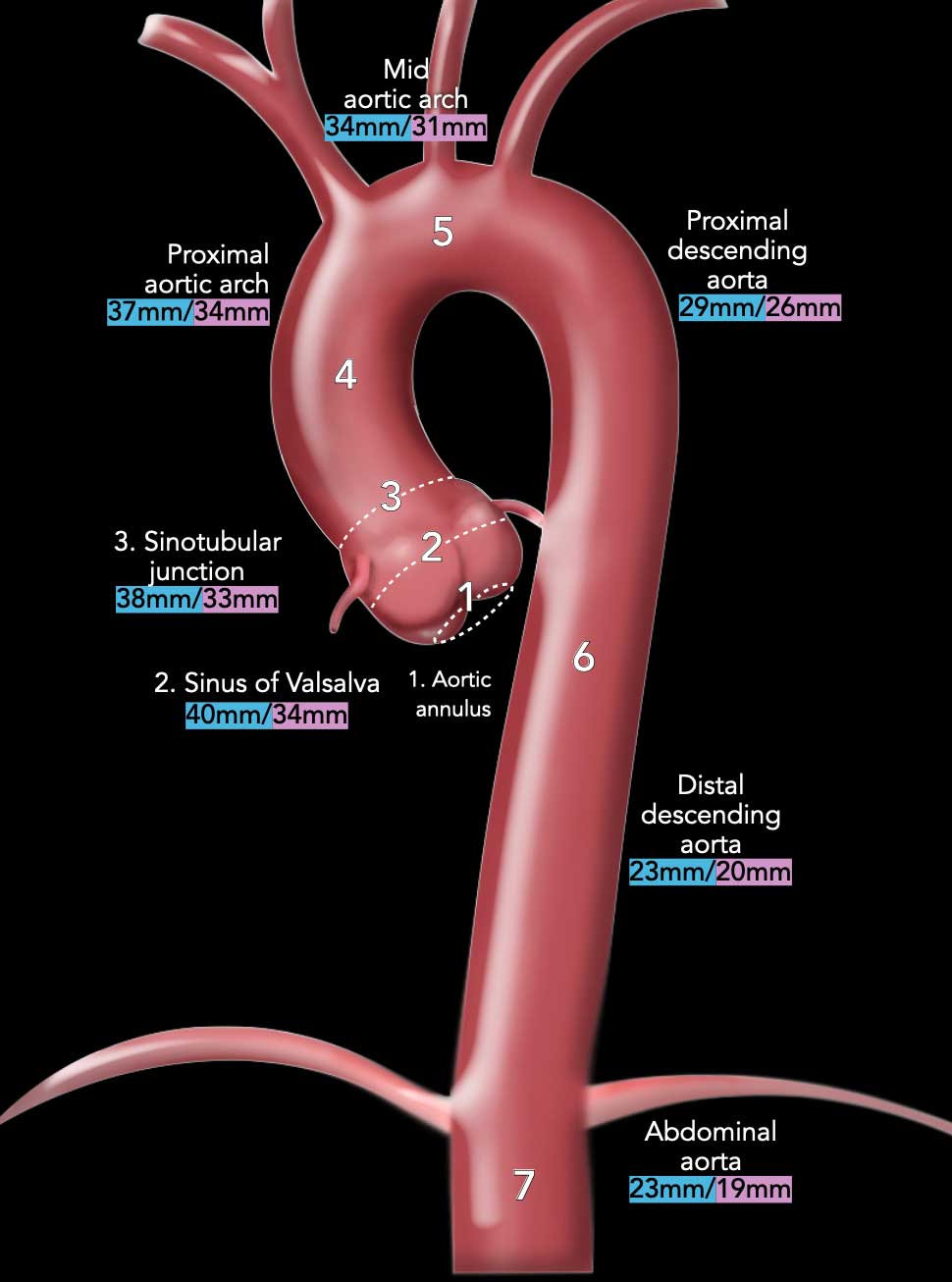
When evaluating the thoracic aorta, measurements are typically obtained at seven standard anatomical levels, though some protocols may include additional points for enhanced precision.
The aortic root is assessed at three distinct levels:
- Aortic annulus
- Sinuses of Valsalva
- Sinotubular junction
Further measurements may be taken at the following levels:
- Ascending aorta
- Proximal aortic arch
- Mid aortic arch
- Descending aorta (with some protocols including two separate measurements)
- Abdominal aorta
Simplified reference values for these measurements are
provided in the table for men (in blue) and women (in pink), not accounting for
body size.
These provide a rough estimate for what constitutes a normal diameter and what
doesn’t.
Measurements
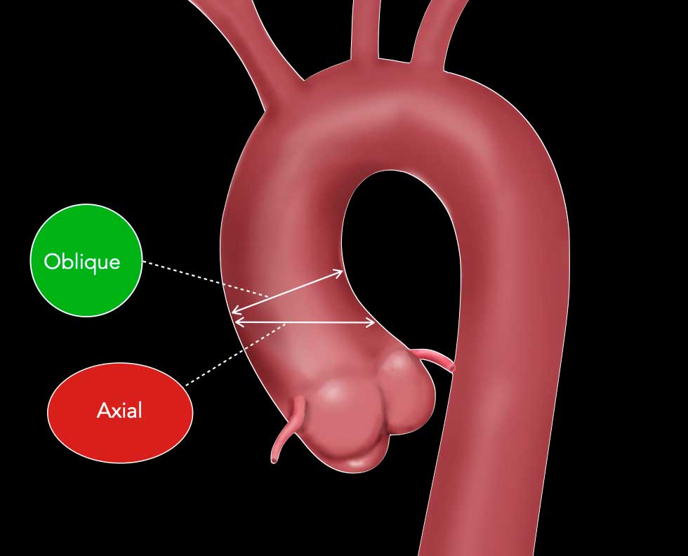
Double-oblique method
The thoracic aorta follows a curved, oblique path.
Axial slices perpendicular to the long axis of the body do not align with the true cross-section of the aorta, leading to elliptical or distorted views rather than circular ones (red ellipse).
The double-oblique method provides the true maximal diameter of the thoracic aorta by aligning the imaging plane perpendicular to the long axis of the aorta.
Technique
- First Oblique Plane: The aorta is visualized in a sagittal or coronal oblique plane to identify its long axis.
- Second Oblique Plane: A perpendicular plane is then created at the level of interest (e.g., ascending aorta, aortic arch, or descending aorta). This plane is orthogonal to the long axis of the aorta, ensuring a true cross-sectional view.
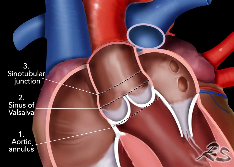
Aortic root
- Aortic annulus: a virtual plane where the valve leaflets (cusps) attach. It is the narrowest portion of the aortic root and the standard measurement used in echocardiography and CT for prosthesis sizing.
- Sinus of Valsalva: three pouches formed by the aortic wall at the level of the aortic valve cusps. They create swirling blood flow, that aid in the efficient closure of the aortic valves.The right coronary artery typically originates from the right sinus of Valsalva, and the left coronary artery originates from the left sinus. The remaining sinus is called the non-coronary sinus.
- Sinotubular junction: the most distal part of the aortic root, marking the boundary between sinus of Valsalva and ascending aorta.
Aortic annulus
In CT imaging, the ‘aortic annular plane’ or ‘hinge point plane’ is typically measured on the slice just below the basal insertion points (nadirs) of the aortic valve cusps (see video).
Make sure that you see all three insertion points in one plane.
Measuring at this level at 20-40% of the cardiac cycle ensures consistency for prosthetic valve sizing (e.g. TAVR/TAVI).
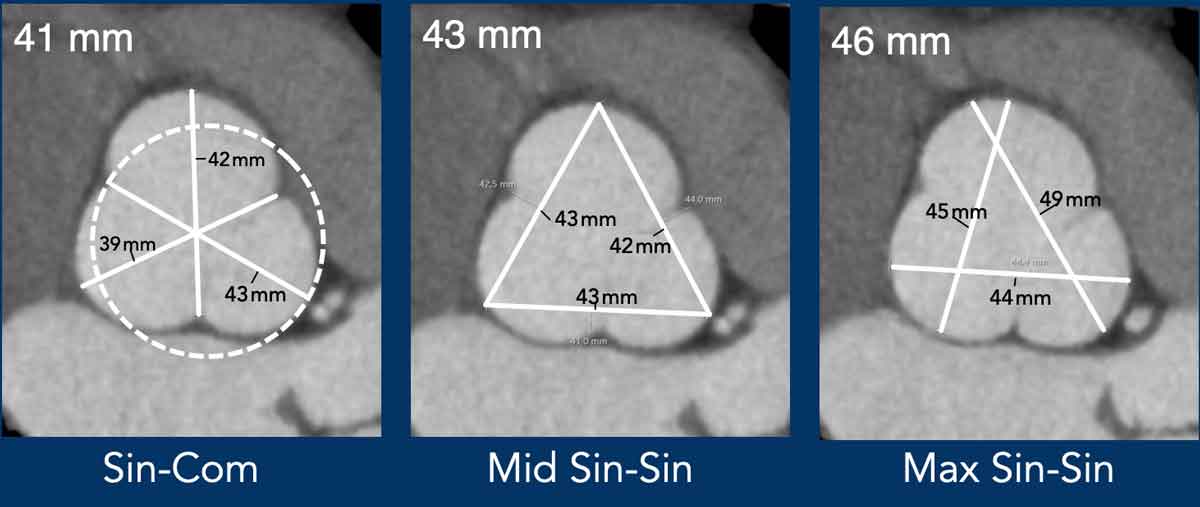
Sinus of Valsalva
The diameter is measured at the
level of maximal sinus bulging, identified by scrolling upward from the
annular plane to the widest point
(referring to the other orientations).
Make sure you do not include the coronary orifices in your measurements.
Measurement Methods
- Sinus-to-Commissure
A line is drawn from the deepest point of one sinus perpendicularly through the center to the opposite commissure. This outdated practice yields a precise but importantly smaller dimension, underestimating the true aortic root diameter (and might as such skew management). - Mid-Sinus to Mid-Sinus
A line connects the centerpoints of opposite sinuses of Valsalva. - Maximum
Sinus-to-Sinus
A line is drawn representing the widest dimension from one sinus to an adjacent sinus. It helps rotating your image to avoid oblique measurements between sinuses. The average of the three sinus-to-sinus dimensions should be calculated, or the largest dimension can be reported.
Note: These methods produce different results.
We prefer the maximum sinus-to-sinus method.
Every institutions should standardize and clearly document their measurement approach.
Video of Root Aneurysms
Thoracic aortic
dilatation most frequently occurs at the level of the ascending aorta.
Aortic root phenotype is defined by dilation of the sinuses of Valsalva exceeding the diameter of the tubular ascending aorta. This morphology is frequently associated with a bicuspid aortic valve.
It is regarded as a risk marker and lowers the threshold for surgical intervention.
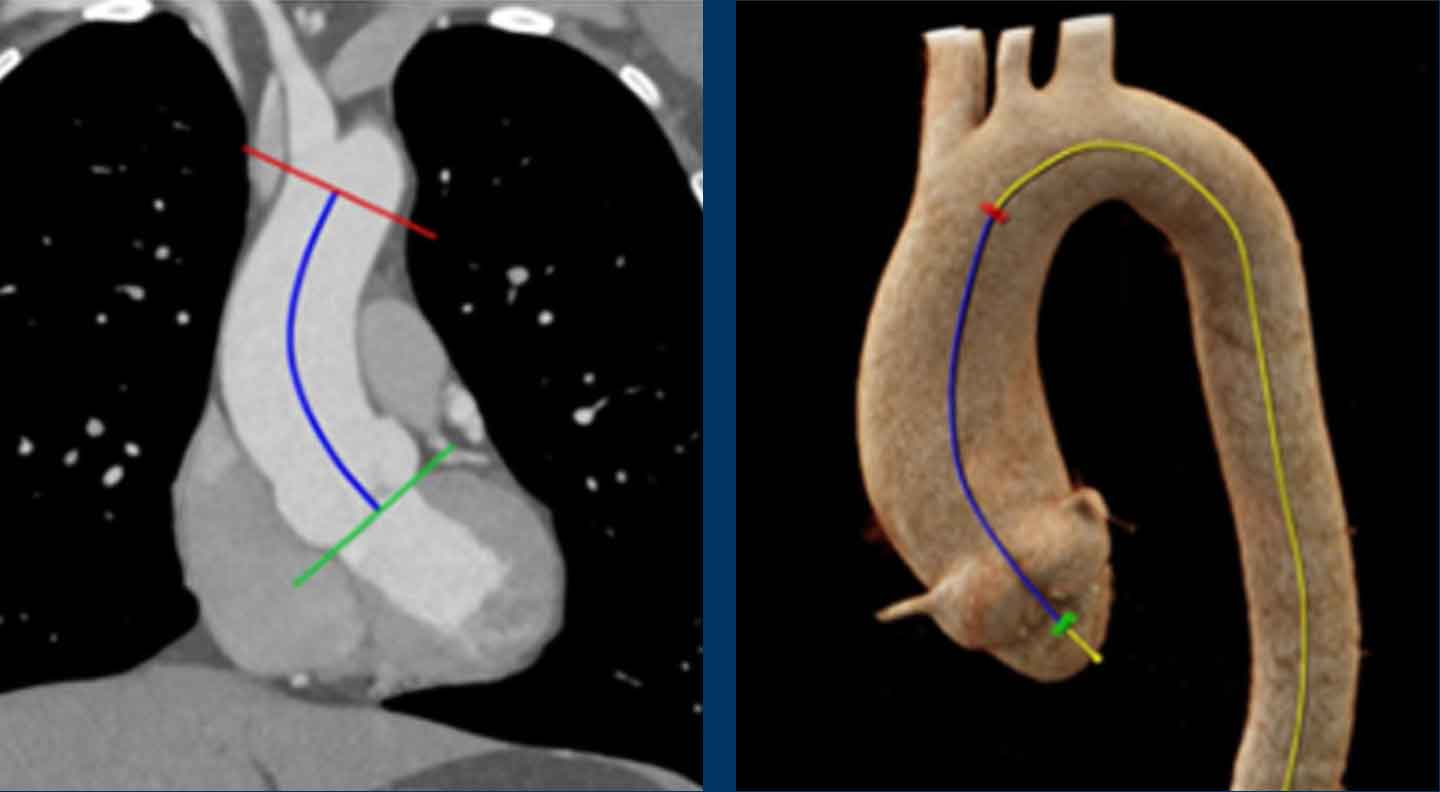
Elongation of Ascending Aorta
Similar to dilation in the transverse plane, longitudinal elongation of the thoracic aorta is associated with an elevated risk of acute complications.
An ascending aortic length exceeding 11 cm — measured from the annulus to the origin of the brachiocephalic artery — serves as a cut-off value for increased risk.
MPR measurements
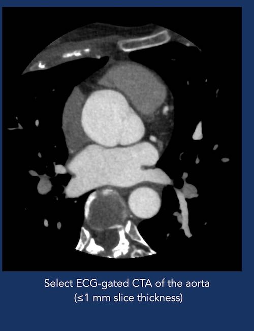
Scroll through the images to see how to measure the thoracic aorta using MPR reconstructions.