Femoroacetabular impingement syndrome
Loes Schiphouwer¹, Adam Weir² and Robin Smithuis³
¹ Haaglanden Medical Center The Hague, ² Erasmus Medical Center Rotterdam and Sport medicine and exercise clinic Haarlem (SBK), and ³ Alrijne hospital in Leiden, the Netherlands
Femoroacetabular
(FAI) impingement syndrome is a
condition characterized by abnormal contact between the femoral head and the
acetabulum, resulting in labral and cartilage damage and causing hip pain,
particularly in young, active individuals.
FAI syndrome is associated with early onset of hip joint osteoarthritis.
The term femoroacetabular impingement syndrome should only be applied to patients with a clinical disorder defined by the triad of:
- Symptoms in young individuals
- Clinical signs at physical examination
- Characteristic radiological findings
This article will focus on the imaging findings associated with FAI syndrome.
Introduction
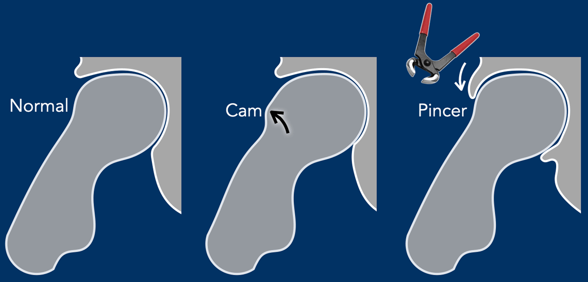
The hip joint is a ball-and-socket joint.
The femoral head, covered with cartilage, serves as the "ball" of the hip joint, while the acetabulum, deepened by the cartilage rim and labrum, functions as the "socket."
With a normal hip joint morphology, there is good congruence during all types of hip movements. If a hip with a normal morphology is moved into supraphysiological end range of motion then even a normal hip may impinge.
The joint congruence can be influenced by bony overgrowth on the femoral side (Cam morphology) or by an extension of the acetabulum over the femoral head, resulting in excessive coverage (Pincer morphology).
Cam and Pincer video
This video demonstrates how Cam and Pincer morphologies can lead to hip impingement.
In this video, only the osseous structures are depicted. However, it is important to understand that impingement also affects the labrum and cartilage rim, potentially resulting in labral tears and focal cartilage loss.

It is important to realize that in order to make the diagnosis of femoroacetabular impingement (FAI) syndrome, patients must present with a combination of the following: symptoms of pain and sometimes reduced motion of the hip, positive clinical signs on testing the hip joint on physical examination, and typical imaging findings.
Since each of these findings alone is non-specific, the diagnosis of FAI syndrome requires their combination. It is crucial to note that an asymptomatic FAI based on imaging alone does not exist. Imaging depicts the morphology of the hip
In Cam morphology, the femoral head has an abnormal shape, often appearing more oval than spherical, which causes abnormal contact with the acetabulum.
This can lead to cartilage damage and hip pain, particularly during
activities involving hip rotation or flexion.
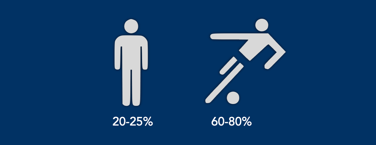
Cam morphology is especially prevalent in healthy athletes.
In studies of professional football players without pain, Cam
morphology was observed in 60–80% of participants, compared to 20–25% in control
populations.
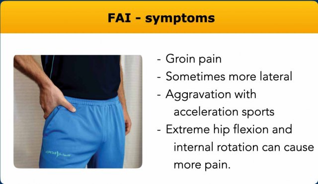
Symptoms
Symptoms of the FAI-syndrome are moderate to marked ventral hip or groin
pain.
The patients often indicate the location of the pain by forming a “c-sign” with their
hand, as it is located deep within the hip.
There is aggravation of the complaints with multi directional sports as
well as squatting, climbing stairs and prolonged sitting.
Moving the hip into deeper hip flexion and internal rotation can increase
the pain.
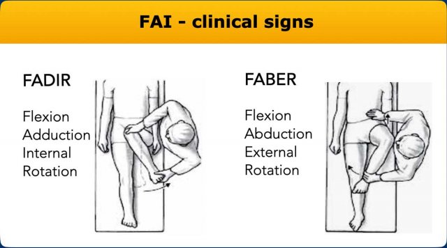
Clinical signs
Hip impingement tests usually reproduce the patient's typical pain.
There is often a limited range of hip motion, typically restricted internal rotation in flexion, especially if you compare to the contralateral side.
The FABER test is flexion combined with abduction and external rotation-
this test can sometimes reproduce the pain.
The most used test is the FADIR test with flexion, adduction and
internal rotation.
These tests are sensitive but not very specific and thus the typical
symptoms and imaging findings should also be present to make the diagnosis of
FAI syndrome.
AP and Dunn view
AP view
The anteroposterior (AP) view of the pelvis plays a key role in identifying bony abnormalities, evaluating joint space, measuring critical angles, and determining the overall alignment of the pelvis, which all contribute to the diagnosis of femoroacetabular impingement.
However, it is often used in combination with other imaging views for a more comprehensive evaluation.
Dunn view
The
Dunn view is an oblique view that
helps to visualize the head-neck
junction in a manner that is not typically seen in standard views like the anteroposterior
(AP) or lateral views.It
is performed by having the patient lie on their back with their affected
hip flexed at about 45° and abducted at 20° to 30° with neutral rotation.
This
positioning allows for a clearer view of the anterosuperior
aspect of the head-neck junction, which is often
difficult to assess on standard views and is the most common location of the
cam morphology.
Images
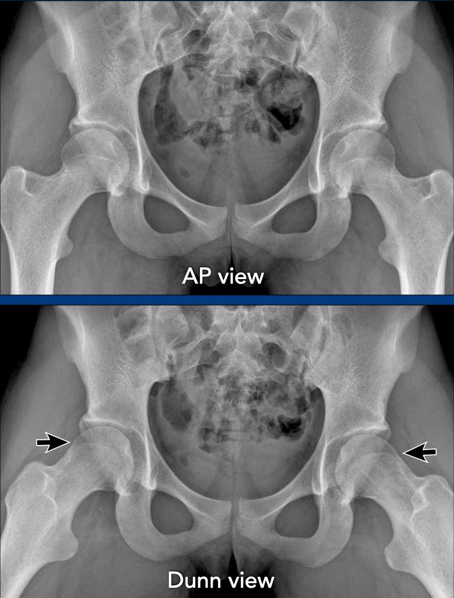
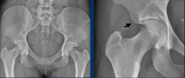
A 20-year-old gymnast with hip complaints.
On the AP view the femoral head-neck region looks normal.
On the Dunn view there is Cam morphology on both sides (arrows).
WEA and CEA measurement (video)
This video shows you how to measure the W-CEA and L-CEA angles. Click on the video for a larger view.
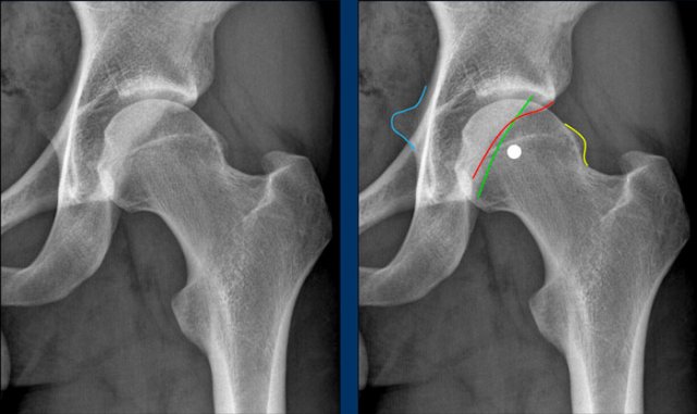
W-CEA
The Wiberg-CEA is the angle between a vertical line through the femoral head center and a line to the lateral margin of the acetabular sourcil, which is the weight bearing and dense area of the superior acetabulum.
It represents the for FAI more relevant anterosuperior coverage.
L-CEA
The lateral CEA is measured to the far lateral acetabular margin and represents the superolateral coverage.
Sometimes these measurements coincide.
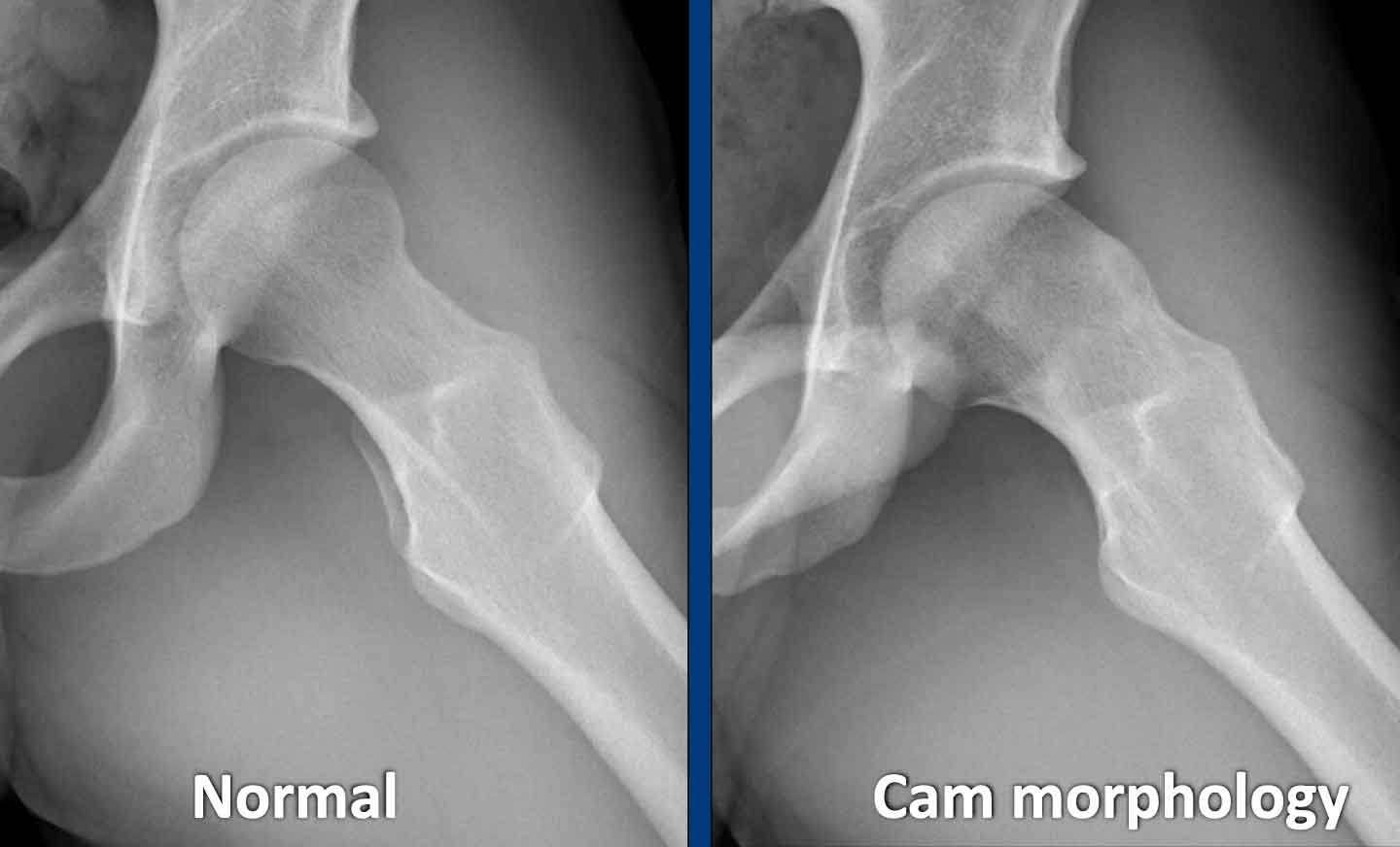
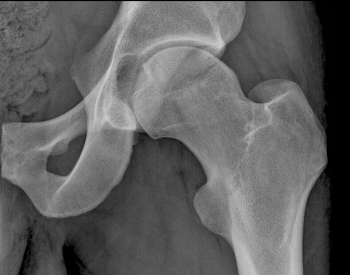
Cam morphology

Cam morphology refers to a benign bony prominence that develops at the
femoral head-neck junction of the hip often in combination with an osseous
asphericity of the femoral head.
This morphology develops during adolescence and does not appear after
the femoral epiphysis has matured. It causes the femoral head and neck to impinge against the
acetabulum leading to damage of the labrum and cartilage which can cause hip
pain, particularly with activities that involve hip rotation or flexion.
There are multiple international
consensus statements that advise using the term Cam morphology. Terms such as Cam lesion, bony bump, or
pistol-grip deformity should be avoided, as it may also be present in many
asymptomatic athletes.
When observing a bony bump or osseous convexity, as seen in the
illustration, it can be referred to as Cam morphology.
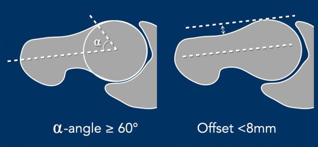
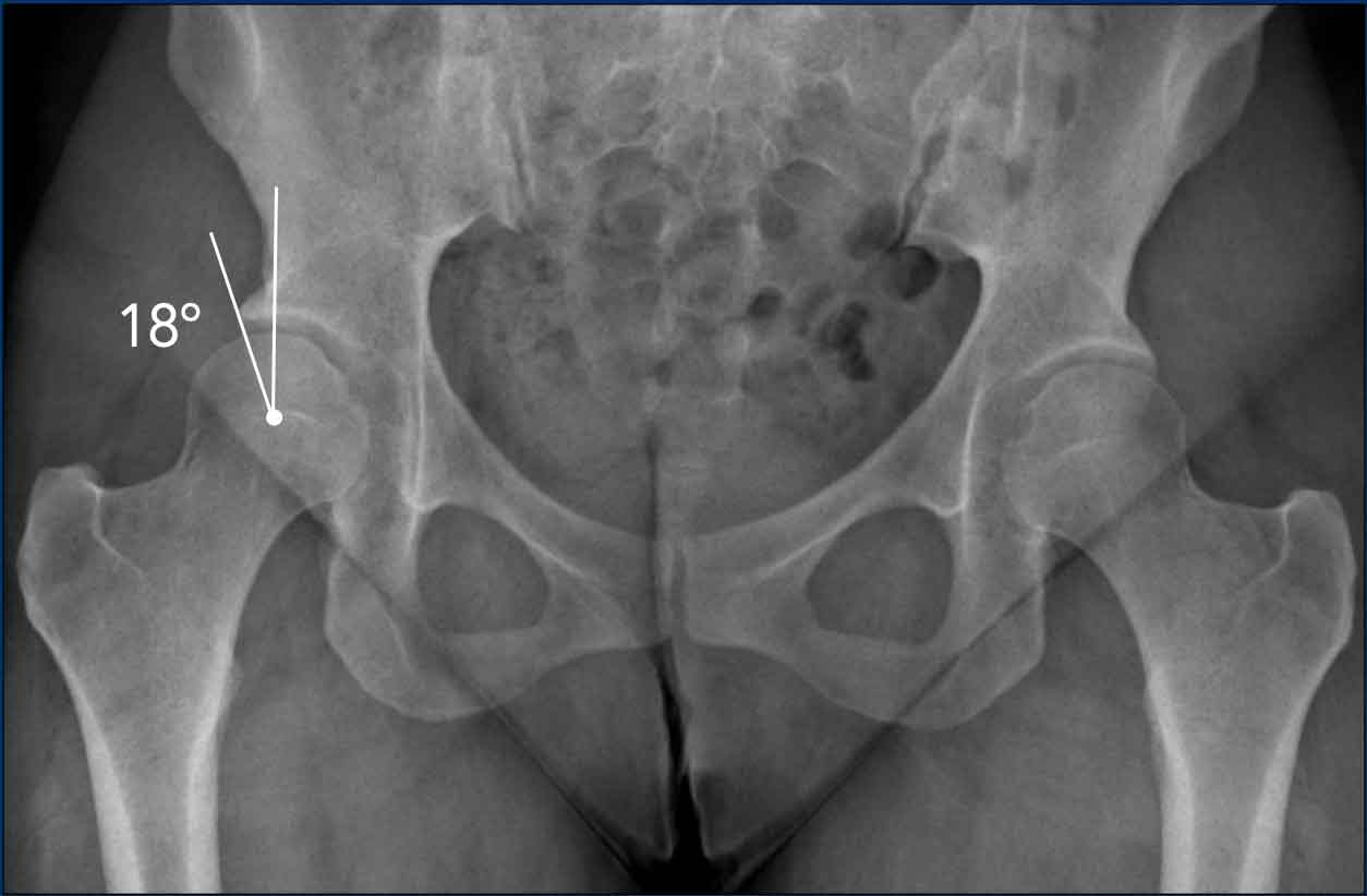
Alpha angle
The
alpha angle is measured by drawing a circle through the femoral head, a line
from the center of the femoral neck to the center of the femoral head, and then
a line from the center of the femoral head to where the femoral head-neck
junction intersects the circle.
If the angle is 60 degrees or more, it can be classified as Cam morphology.
This measurement can be used both on conventional imaging as well as
cross-sectional imaging - with cross-sectional imaging being more sensitive.
The threshold has been increased recently to 60 degrees to make it more
specific.
Offset
Additionally, the offset can also be measured on cross-sectional imaging (CT or
MRI).
For this, a line is drawn on the anterior side of the femoral head, parallel to
the femoral neck.
If the distance between this line and the anterior side of the femoral neck is
less than 8 mm, it can be classified as Cam morphology.
Alpha angle measurement
The normal hip shows an alpha-angle less than 60º.
A hip with the Cam morphology has an alpha-angle greater than 60º.
Of course, when the Cam morphology is as pronounced as in this case, these measurements are unnecessary in clinical practice.
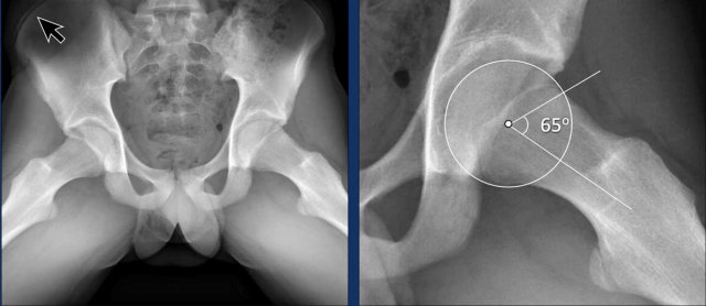
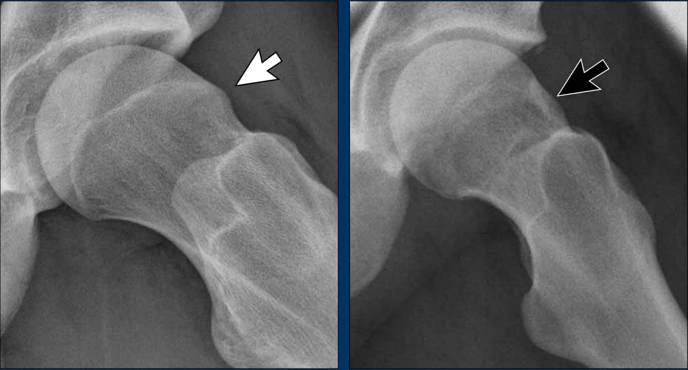
This is a more subtle case of Cam morphology in a young male.
Notice that the iliac crest growth plate is not yet closed (arrow).
Images
Two examples of Cam morphology.
Pincer morphology
Pincer morphology is characterized by an abnormal orientation or abnormal shape of the acetabulum, such as acetabular retroversion or acetabular protrusion. This results in excessive or abnormal coverage of the femoral head, which can lead to impingement.
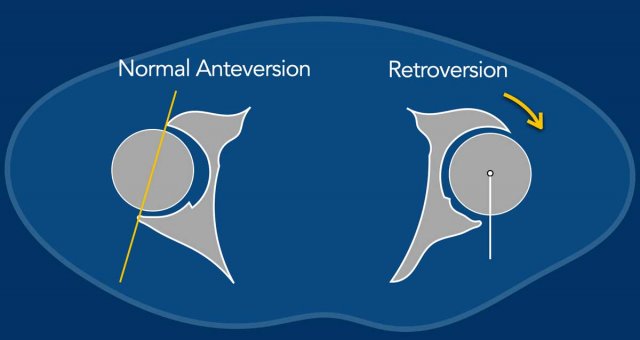
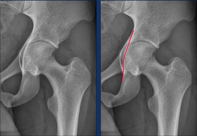
Acetabular Retroversion
The normal acetabulum has an anteverted orientation (figure), meaning it is more open on the anterior side compared to the posterior side. On radiographs, the anterior wall projects medially to the posterior wall.
In acetabular retroversion, the acetabulum is abnormally oriented,
leading to excessive coverage of the femoral head on the anterior side, which
can result in impingement during flexion and internal rotation. In these cases
patients often have a great degree of external rotation on clinical
examination. In addition the ischial spine will project more medially (ischial
spine sign).
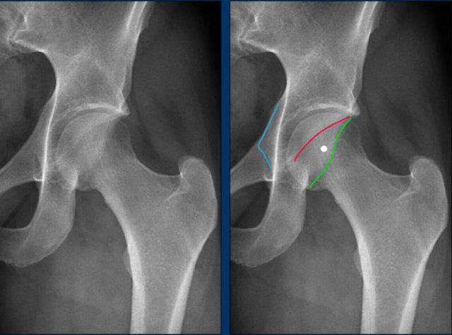
Normal anteversion of the acetabulum
This is a detail of the normal left hip on an AP Pelvis X-ray.
These measurements should always be assessed on a pelvic x-ray and not
one centered on a single hip.
As a result of the normal anteverted orientation of the acetabulum, the border of the anterior wall (red line) projects medially to the posterior wall (green line) , i.e. no cross over.
The posterior wall projects laterally to the center of the femoral head, i.e. no posterior wall sign.
The ischial spine (blue line) does not project medially, i.e. no ischial spine sign.
Acetabular Retroversion
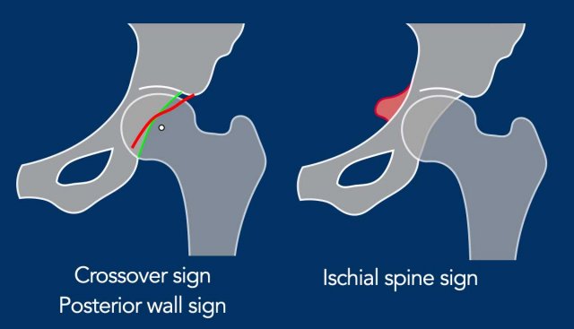
On radiographs, signs of acetabular retroversion include:
Cross-over sign - the anterior wall (especially the superior portion) projecting laterally to the posterior wall.
Posterior wall sign - the posterior wall projecting medially to the center of the femoral head.
Ischial spine sign - the ischial spine projecting more medially than normal.
If only the cross-over sign is positive, it is referred to as a focal (anterosuperior) pincer.
When
both the posterior wall sign and the ischial spine sign are also positive, it
is classified as a global pincer/acetabular retroversion.
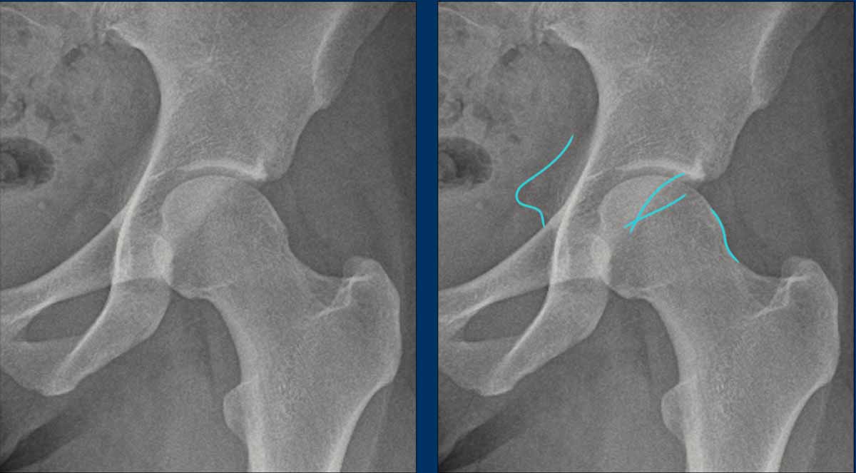
This is a detail of an AP Pelvis X-ray.
First study the image.
Then scroll to the next image.
Findings
- Cross-over sign - the superior portion of the anterior acetabular wall (red line) extends laterally beyond the posterior acetabular wall.
- Posterior wall sign - the posterior wall projects medial to the center of the femoral head.
- Ischial spine sign - the ischial spine projects more medially than normal (blue line).
These findings are consistent with a global pincer morphology due to acetabular retroversion.
Notice that there is also some cam-morphology.
This is another example of pincer morphology on an image zoomed in from an AP pelvic radiograph.
There is a subtle cross over sign.
In addition there is a posterior wall sign and ischial spine sign.
Notice that there is also cam-morphology (yellow line).
This is another example of pincer and cam morphology on an AP pelvic radiograph.
Scroll through the images.
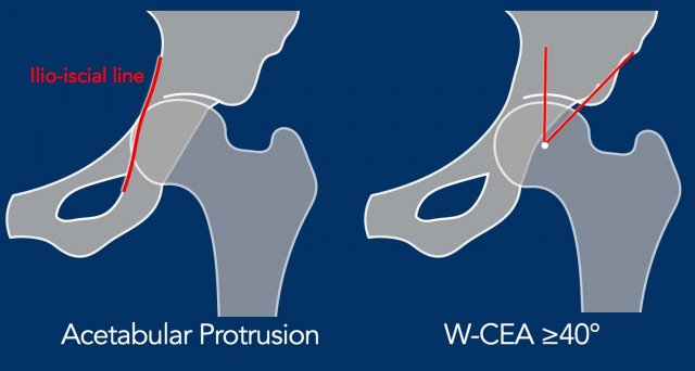
Acetabular protrusion
Ilio-ischial line
In acetabular protrusion, the acetabulum is too deep with inward displacement and global overcoverage of the femoral head. If the medial border of the femoral head extends beyond the ilio-ischial line, this is referred to as acetabular protrusion. This is an uncommon
finding in athletes.
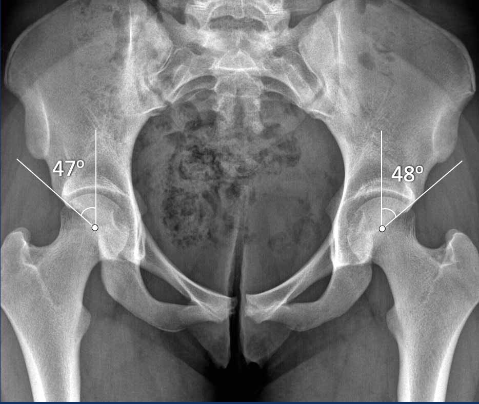
CEA angle
When
measuring the center-edge angle (CEA), the lateral boundary of the
anterosuperior wall/lateral border of the acetabular sourcil should be used, also known as the lateral center edge angle of Wiberg/W-CEA.
This can be recognized on an AP x-ray by using the lateral margin of the subchondral
sclerotic zone of the superior acetabulum.
When using the most lateral border
of the acetabulum (which is usually reflective of the border of the superolateral
acetabulum rather than the anterosuperior border), or the L-CEA, the coverage might
be overestimated.
If there is a retroversion of the (superior) acetabulum, the
W-CEA and L-CEA can be the same.
If this angle is equal to or greater than 40 degrees, it is classified as global pincer with overcoverage of the femoral head.
Acetabular dysplasia
Unrecognized hip acetabular dysplasia is sometimes clinically mistaken
for FAI-related complaints.
It is important to also report any potential undercoverage or dysplasia.
(i.e. L-CEA < 20 degrees).
Many consider a L-CEA between 20 and 25 degrees borderline dysplasia.
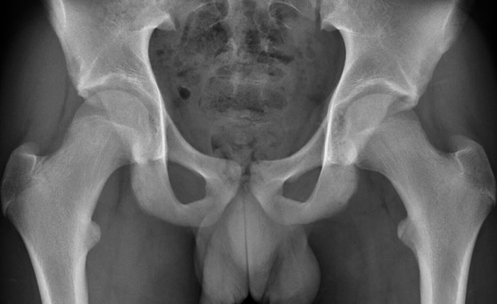
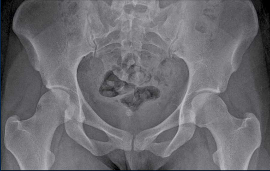
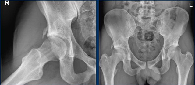
This image is of a 23-year-old male presenting with hip-related complaints.
Image
This is a case of acetabular protrusion, where the femoral head just crosses
the ilioischial line.
Image
Isolated pincer on both sides (only AP view available) with increased
W-CEA and ischial spine sign in a 16 year old female patient, who presented
with hip-related complaints.
Treatment
Conservative therapy
In most cases, an initial attempt is made to improve symptoms through
conservative therapy. This treatment involves providing insight into the origin
of the symptoms, advising the avoidance of extreme postures. It often helps to
sit in less deep hip flexion positions. Exercises to strengthen the muscles around the hip joint are used. Attention can be given to the
position of the legs and pelvis during repetitive sports motions.
Arthroscopic surgery
If these measures prove ineffective, surgical intervention may be
considered. Prior to surgery, an MR arthrography will often be performed to
assess the condition of the labrum and rule out other possible pathologies,
such as severe chondral damage or idiopathic bone marrow edema, among others.
Small labral tears are typically left untreated, while larger unstable tears
may be either excised or repaired. During surgery the excessive bone of the cam
morphology on the femoral side of the acetabular rim can be removed. Rehabilitation
following surgery takes a long time and it will often take at least 9 months to
return to high level sports.
There are randomized trials showing that surgery is slightly more effective than conservative therapy. Many athletes who undergo treatment still have some symptoms in the long term and not all are able to return to their previous level of activity.
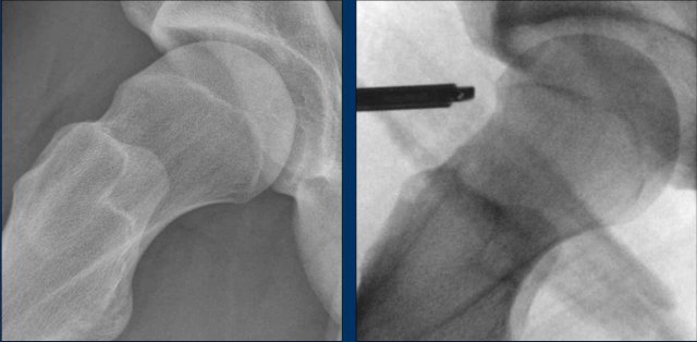
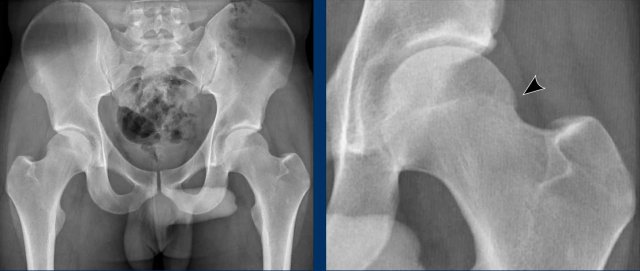
Images
In a patient with Cam morphology, the effect of reshaping of the femoral
head-neck junction is seen (arrow).
Examples of FAI
Combination of Cam and Pincer morphology
These images are from a 28-year-old woman who participates in CrossFit at a professional level. She has been experiencing complaints in the left hip region, specifically on the anterior side, for the past year. The symptoms worsen during squats and hip flexion.
Upon examination, there is reduced hip flexion (painful at 120°) and limited rotation (restricted to 20° and painful).
First study the radiograph.
Then continue with the detailed views...
Although the findings are subtle, there is evidence of Pincer morphology, indicated by the crossover sign and ischial spine sign.
The retroversion of the acetabulum results in increased prominence of the ischial spine and leads to the superior portion of the anterior wall of the acetabulum overlapping the posterior wall on imaging.
Furthermore, there is a mild indication of Cam morphology.
Continue with the MR-arthrogram...
Subsequently, MR arthrography was performed, revealing a labral tear in the anterosuperior region.
Initially, the patient was treated conservatively with physiotherapy, but this did not lead to satisfactory results.
An arthroscopic procedure was then performed, involving a reshaping of the femoral head-neck junction (arrow), which resulted in significant improvement.
Cam and osteoarthritis
This is a 27-year-old man who developed deep ventral groin pain seven
years ago while squatting.
Since then, he has stopped playing football but has continued to
experience the pain.
Images
The radiographs reveal a bony prominence on the femoral head/neck
consistent with cam morphology.
Continue with the MRI...
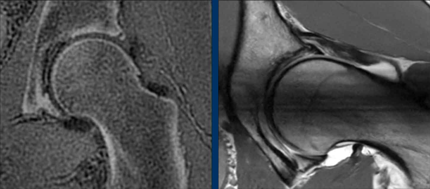
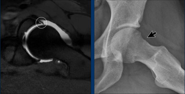
Images
The
image on the left is a so-called bone/zero TE (ZTE) MRI.
It is a special MRI acquisition to study the bones.
The images look quite similar to CT images and can be made as part of a
MR-arthrogram of the hip.
It shows a small osteophyte (black arrow).
Scroll through the images of the MR-arthrogram on the right side.
There is a cartilage defect on the acetabular side (yellow arrow).
Conclusion
Since there is already a subtle osteoarthritis in this patient, he is
not a candidate for an arthroscopic treatment.
He was treated conservatively with physiotherapy, exercise therapy,
education and activity modification.
Subspine impingement
Subspine Impingement is a condition in which the anterior inferior iliac spine, also known as the subspine, impinges upon the femoral neck or head, especially during activities requiring deep hip flexion or rotation, such as sports or yoga.
These images are of a 23-year-old woman with a history of rheumatoid arthritis.
She presented with progressive pain in the right hip, particularly during flexion movements while practicing yoga.
Images
There is a variant with a low position of the inferior iliac spine (arrow).
This is another example of subspine impingement in a 28-year-old amateur
football player who had acute pain in the
right ventral hip region during sprinting eight months ago.
Since then, he has experienced progressive hip complaints.
Physical examination reveals significantly reduced flexion, as well as
decreased internal and external rotation.
Image
There is a typical calcification following an injury of the rectus
femoris tendon around the insertion at the anterior inferior iliac spine.
This has resulted in bony overgrowth, causing impingement on the femoral
head/neck region.
Surgical
removal of the ossification was performed.