Temporal Bone Anatomy 1.0
Erik Beek and Robin Smithuis
Radiology department of the University Medical Centre of Utrecht and the Rijnland Hospital, Leiderdorp, the Netherlands
Publicationdate
Updated version: 21-2-2007
In this review we present the normal coronal and axial anatomy of the temporal bone.
Learn the anatomy by scrolling through the images.
Temporal bone
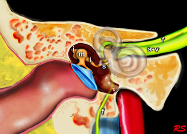
The middle ear consists of the tympanic cavity and the antrum.
The antrum is a large aircell superior and posterior to the tympanic cavity and connected to the tympanic cavity via the aditus ad antrum.
The epitympanum or attic is the upper portion of the tympanic cavity above the tympanic membrane, and contains the head of the malleus and the body of the incus.
The tympanic membrane, the malleus, incus and stapes transfer soundwaves to the stapes footplate, which is attached to the base of the cochlea in the oval window.
Axial anatomy
Scroll through the axial anatomy from inferior to superior
- A = Antrum
- f = facial nerve
- genu = first genu of facial nerve at the geniculate ganglion
- IAC = internal auditory canal
- long crus = long crus of the incus
- short crus = short crus of the incus
- U = utricle of vestibule
Axial anatomy from inferior to superior
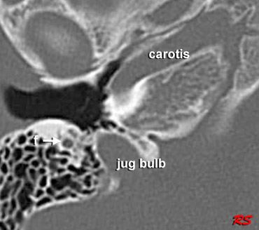
At the most inferior level we see the facial nerve passing inferiorly to finally reach the stylomastoid foramen (not shown in this image).
The carotid artery is shown within the carotid canal.
Also at this level is the top of the jugular bulb.
Tympanic membrane
The manubrium of the malleus (yellow arrow) is connected to the tympanic membrane.
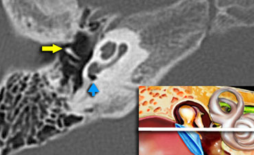 Malleus (yellow arow). Round window (blue arrow)
At this level we can see the manubrium of the malleus (yellow arow) anterior to the long process of the incus.
The round window is indicated by the blue arrow.
The round window dissipates the pressure generated by the fluid vibrations within the cochlea and thus serves as a release valve.
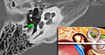 Stapes (green arrow) is seen connecting to the oval window.
Stapes
The base of the stapes rocks in and out against the oval window.
The vibrations are transmitted via the endolymph to the hair cells of the organ of Corti of the cochlea.
Cochlea
Within the cochlea the movement of the hair cells convert the sound-vibrations into nerve impulses, that travel over the cochlear nerve to the auditory cortex of the brain, which interprets the impulses as sound. .
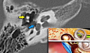
The head of the malleus is seen anterior to the head of the incus (yellow arrow).

Tympanic segment of the facial nerve
In this image at the level of the internal auditory canal, the tympanic segment of the facial nerve is seen just medial and parallel to the wall of the epitympanum.
The head of the malleus (yellow arrow) is seen anterior to the head and the short process of the incus.

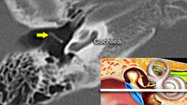
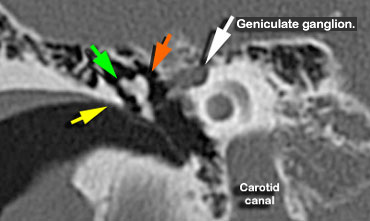
Geniculate ganglion of the facial nerve
At this level the aditus ad antrum is seen. This is the connection between the tympanic cavity and the antrum.
The labyrinthine segment of the facial nerve coming from the internal auditory canal angles sharply forward, nearly at right angles to the long axis of the petrous bone, to reach the geniculate ganglion.
At the ganglion the facial nerve makes a U-turn (first genu of the facial nerve) to run posteriorly as the tympanic segment along the medial wall of the epitympanum.

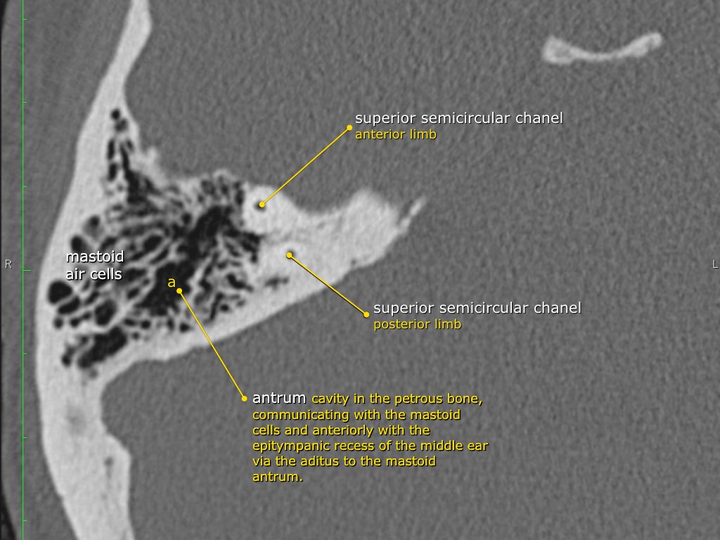
Antrum
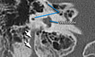
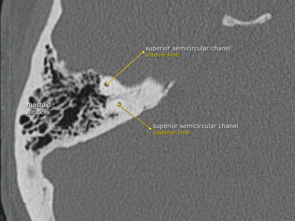
At this level the antrum is seen surrounded by smaller mastoid aircells just lateral to the superior semicircular canals .
The three semicircular canals lie perpendicular to each other to sense acceleration and deceleration movements in each of the 3 spatial planes.
Static head position is sensed by the vestibule, which contains the position hair cells.
Different head positions produce different gravity effects by small calcium carbonate particles (otoliths) on these hair cells.
Coronal anatomy
The petrous bone is positioned in an oblique orientation from posterolateral to anteromedial.
As a result most structures will be sectioned obliquely on coronal images.
The following coronal images go from anterior to posterior.
First we will see the tympanic membrane with the ossicles, followed by the cochlea, antrum and semicircular canalls.
Finally the most posterior image will show the point where the facial nerve exits the temporal bone at the stylomastoid foramen.
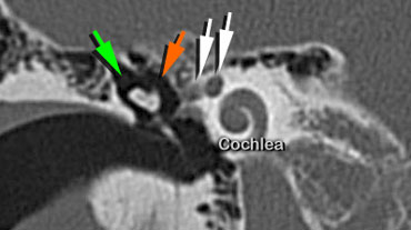
 Scutum (yellow arrow). Head of malleus (orange arrow) is seen medial to the incus (green arrow)
Scutum
The scutum (yellow arrow) is a sharp bony spur formed by the lateral wall of the tympanic cavity and the superior wall of the external auditory canal.
It is usually the first bony structure to erode as a result of a cholesteatoma, that is formed by medial retraction of the pars flaccida of the tympanic membrane into the epitympanum.
If the retraction continues it will result in ossicular destruction.
If the cholesteatoma passes posteriorly through the aditus ad antrum into the mastoid itself, erosion of the tegmen mastoideum, with exposure of the dura and erosion of the lateral semicircular canal with deafness and vertigo, may result.
On the left the most anterior point of the facial nerve is seen (white arrow).
At this point the nerve makes a U-turn.
It is named the genu or geniculum and represents the geniculate ganglion.
The malleus is seen connected to the tympanic membrane and to the incus.
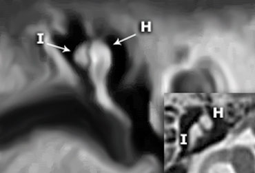 Coronal reconstruction clearly demonstates that the incus (I) is positioned posterolateral to the malleolar head (H).On the axial image the short crus of the incus is seen pointing posterolaterally.
In many illustrations you will see the incus connecting medially to the malleus, but this is not correct.
On the coronal reconstruction on the left it is clearly demonstated that the incus is positioned posterolaterally to the malleolar head.
The long crus of the incus subsequently runs inferomedially to the stapes.
 Head of malleus (orange arrow) is seen medial to the Incus (green arrow)
A coronal image slightly more posteriorly will show the facial nerve twice.
The medial portion is the part that exits the internal auditory canal and runs towards the geniculate ganglion (medial white arrow).
The lateral portion is the part that courses in a posterior direction, coming from the U-turn of the first genu.
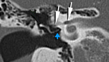 Long crus of the incus is seen connecting to the Stapes (blue arrow).Facial nerve in internal auditory canal and tympanic segment (white arrows).
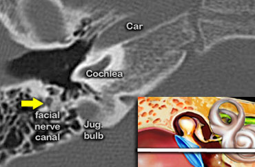
Facial nerve canal
The facial nerve is seen in the internal auditory canal and entering the temporal bone (medial white arrow).
The lateral white arrow represents the tympanic segment of the facial nerve running in the facial canal and curving around the oval window niche.
At this point, the nerve runs in a horizontal plane in a posterior direction superiorly to the oval window .
The incus (orange arrow) is seen connecting to the stapes (blue arrow).

Coronal scan showing the facial nerve (white arrow) above the oval window and below the lateral semicircular canal.
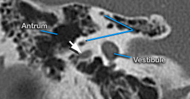
Antrum
The antrum is a large aircell superior and posterior to the tympanic cavity and connected to the tympanic cavity via the aditus ad antrum.
It is surrounded by smaller mastoid aircells.
On this last posterior coronal image the facial nerve assumes a vertical position to exit the petrous bone through the stylomastoid foramen.
Test...
Anatomy
Anatomy