Liver - Segmental Anatomy
Robin Smithuis and Eduard E. de Lange
Radiology Department of the Alrijne Hospital, Leiderdorp, the Netherlands and University of Virginia Health System, Charlottesville, USA.
Publicationdate
The anatomy of the liver can be described using two different aspects: morphological anatomy and functional anatomy.
The traditional morphological anatomy is based on the external appearance of the liver and does not show the internal features of vessels and biliary ducts branching, which are of obvious importance in hepatic surgery.
The French surgeon and anatomist Claude Couinaud was the first to divide the liver into eight functionally indepedent segments allowing resection of segments without damaging other segments.
Segmental anatomy
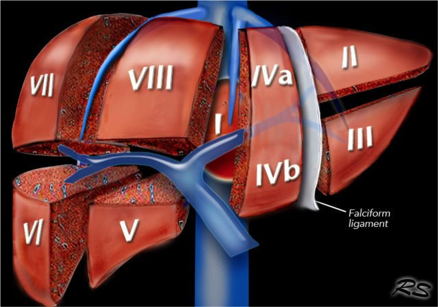 Segmental anatomy according to Couinaud. Click to enlarge.
Couinaud classification
The Couinaud classification of liver anatomy divides the liver into eight functionally indepedent segments.
Each segment has its own vascular inflow, outflow and biliary drainage.
In the centre of each segment there is a branch of the portal vein, hepatic artery and bile duct.
In the periphery of each segment there is vascular outflow through the hepatic veins.
The liver is divided in three vertical planes:
- The plane of the right hepatic vein divides the right lobe into anterior and posterior segments.
- The plane of the middle hepatic vein divides the liver into right and left lobes or right and left hemiliver. This plane runs from the inferior vena cava to the gallbladder fossa.
- The umbilic plane runs from the falciform ligament to the inferior vena cava and divides the left lobe into a medial part, which is segment IV and a lateral part formed by segment II and III. This division is the only vertically oriented plane that is not defined by a hepatic vein.
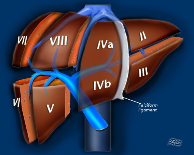
Here another illustration of the functional segmental liver anatomy.
Portal vein
The portal vein divides the liver into upper and lower segments.
The left and right portal veins branch superiorly and inferiorly to project into the center of each segment.
Left hepatic vein
The significance of the left hepatic vein is somewhat controversial. Some authors have shown it to coincide with the umbilical fissure, but in reality the left hepatic vein courses to the lateral to the umbilical fissure [fig].
While some authors have claimed that the division between segments II and III is formed by the transverse plane of the left portal vein, most investigators feel that it is the plane defined by the left hepatic vein.
In actual practice, when a lesion is located within the lateral segment of the left lobe, both Couinaud segments II and III are usually removed based on the plane formed by the umbilical fissure (i.e. left lateral segmentectomy).
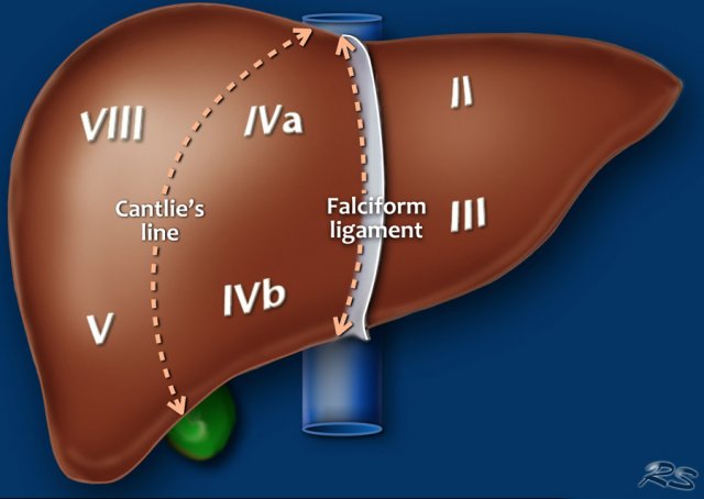 On a frontal view of the liver the posteriorly located segments VI and VII are not visible.
The illustration above is a schematic presentation of the liver segments.
In reality however the proportions are different.
On a normal frontal view the segments VI and VII are not visible because they are located more posteriorly.
The right border of the liver is formed by segment V and VIII.
Although segment IV is part of the left hemiliver, it is situated more to the right.
Couinaud divided the liver into a functional left and right liver by a main portal scissurae containing the middle hepatic vein.
This is known as Cantlie's line.
Cantlie's line runs from the middle of the gallbladder fossa anteriorly to the inferior vena cava posteriorly.
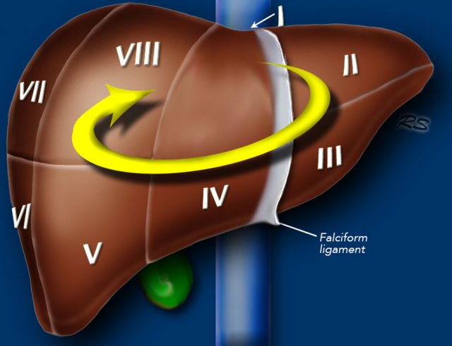 Clockwise numbering of the segments
Segments numbering
There are eight liver segments.
Segment IV is sometimes divided into segment IVa and IVb according to Bismuth.
The numbering of the segments is in a clockwise manner.
Segment I (the caudate lobe) is located posteriorly.
It is not visible on a frontal view.
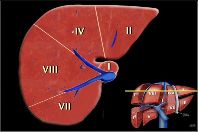 Image at the level of the superior liver segments.
Transverse anatomy
This figure is a transverse image through the superior liver segments, that are divided by the right and middle hepatic veins and the falciform ligament.
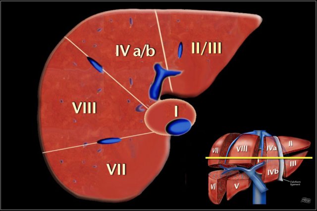 Image at the level of the left portal vein.
This is a transverse image at the level of the left portal vein.
At this level the left portal vein divides the left lobe into the superior segments (II and IVa) and the inferior segments (III and IVb).
The left portal vein is at a higher level than the right portal vein.
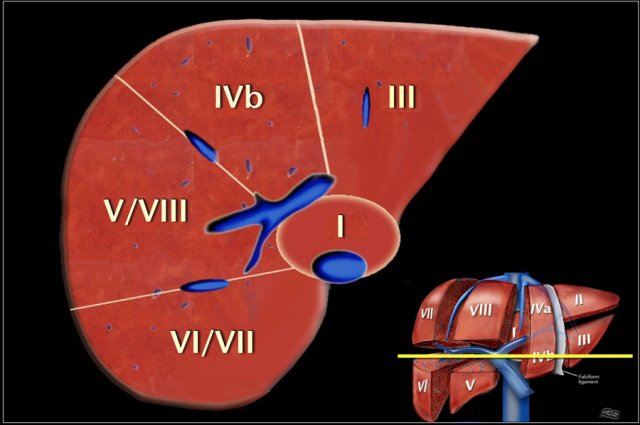 Image at the level of the right portal vein.
This image is at the level of the right portal vein.
At this level the right portal vein divides the right lobe of the liver into superior segments (VII and VIII) and the inferior segments (V and VI).
The level of the right portal vein is inferior to the level of the left portal vein.
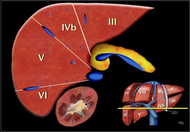 Image at the level of the splenic vein.
At the level of the splenic vein, which is below the level of the right portal vein, only the inferior segments are visible.
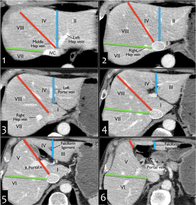
How to separate liver segments on cross sectional imaging
Left liver: lateral(II/III) vs medial segment (IVA/B)
Extrapolate a line along the falciform ligament superiorly to the confluence of the left and middle hepatic veins at the IVC (blue line).
Left vs Right liver: IVA/B vs V/VIII
Extrapolate a line from the gallbladder fossa superiorly along the middle hepatic vein to the IVC (red line).
Right liver: anterior (V/VIII) vs posterior segment (VI/VII)
Extrapolate a line along the right hepatic vein from the IVC inferiorly to the lateral liver margin (green line).
Video of MRI anatomy
 Hypertrophy of caudate lobe in a patient with livercirrhosis. Notice the small lobulated right hemiliver.
Caudate lobe
The caudate lobe or segment I is located posteriorly.
The caudate lobe is anatomically different from other lobes in that it often has direct connections to the IVC through hepatic veins, that are separate from the main hepatic veins.
The caudate lobe may be supplied by both right and left branches of the portal vein.
This CT-image is of a patient with liver cirrhosis with extreme atrophy of the right lobe, normal volume of the left lobe and hypertrophy of the caudate lobe.
Due to a different blood supply the caudate lobe is spared from the disease process and hypertrophied to compensate for the loss of normal liverparenchyma.
Liver surgery
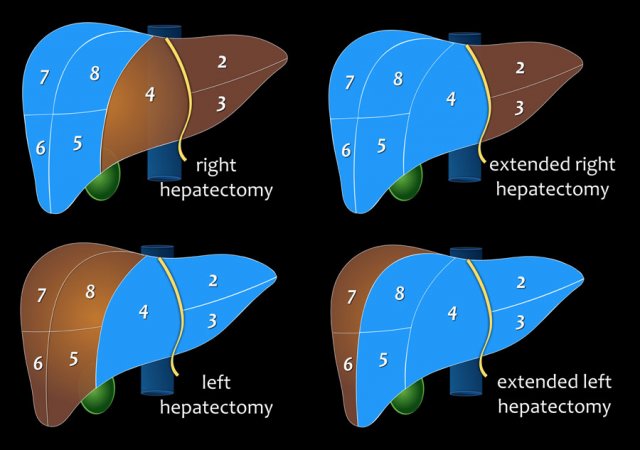
Right hepatectomy
segment V, VI, VII and VIII (± segment I).
Extended Right or right trisectionectomy
segment IV, V, VI, VII and VIII (± segment I).
Left hepatectomy
segment II, III and IV (± segment I).
Extended Left or left trisectionectomy
segment II, III, IV, V and VIII (± segment I).
Many surgeons prefer to use the term "extended" instead of trisectionectomy to indicate that some adjacent tissue of segment 4, or 5/8, as applicable is included rather than the entire segment 4, or 5/8.
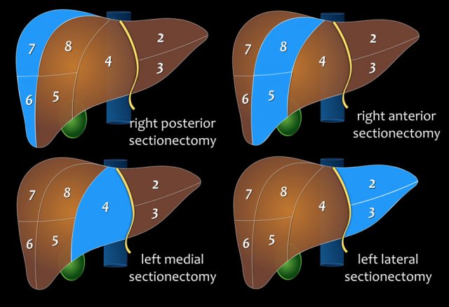
Right posterior sectionectomy
segment VI and VII
Right anterior sectionectomy
segment V and VIII
Left medial sectionectomy
segment IV
Left lateral sectionectomy
segment II and III