Ultrasound of the Breast
Robin Smithuis, Lidy Wijers and Indra Dennert
Alrijne hospital in Leiderdorp - the Netherlands
This article provides a basic understanding of breast-ultrasound.
We will focus on how mobile ultrasound can be used by health workers in regions where other modalities like mammography or MRI are not readily available.
However this article is also a nice introduction for those who do mammography and MRI of the breast.
The videos in the introduction cover most of the material of the article.
Introduction
Video 1
In this video we will discuss:
- Normal anatomy
- Differential diagnosis of a mass in the breast
- Dense glandular tissue
- Cysts
- Fibroadenoms
- Breast cancer
Video 2
In this video we will discuss:
- Many examples of breastcancer
- Intracystic cancer
- Dd benign versus malignant tumor
- Tissue harmonic imaging
- Diagnosis and treatment of an abscess in the breast
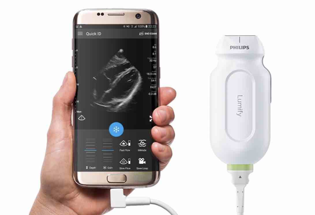 Linear array transducer connected to smart phone
US is mostly used in addition to mammography and MRI.
However it is an excellent primary tool to examine the breasts in symptomatic women who have a lump or localized pain in the breast.
In most cases it can be decided whether we are dealing with a benign condition or breast cancer.
In some cases aspiration can help to differentiate complicated cyst from solid lesions and aspiration can be used to treat abscesses.
Breast ultrasound is not usually done to screen for breast cancer. This is because it is time consuming and you may miss some early signs of cancer like small not-palpable breast cancer or ductal carcinoma in situ which can present as only small calcifications seen on a mammogram.
That said, ultrasound is an extremely fast and powerful tool.
It is safe, cheap and mobile and modern handheld-ultrasound machines consist of only a transducer connected to a mobile phone or tablet.
These systems provide high resolution images for just over one thousand dollars.
We expect that more doctors and health workers will use ultrasound in breastcare when no other modalities like mammography or MRI are affordable or available.
The connection to a mobile phone makes it easy to store images or to share them for a second opinion.
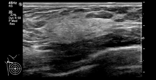
Normal Breast
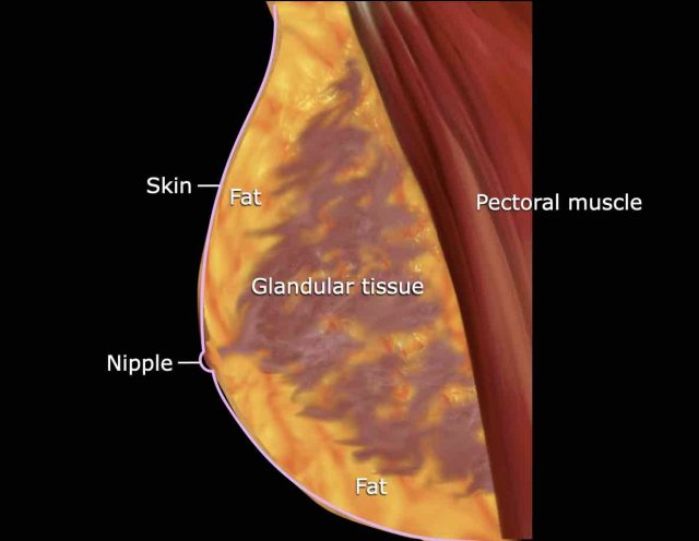
The breast consists of a mixture of fibroglandular and fatty tissue.
The glandular tissue is not evenly spread in the breast.
Usually it is more pronounced in the upper lateral quadrant and it radiates from behind the nipple to more peripheral
The amount of glandular tissue responds to hormonal fluctuation and shows changes during the menstrual cycle.
The amount of glandular tissue decreases with age.
Focal collections of glandular tissue may present as a lump, because glandular tissue is more firm than the surrounding fatty tissue.
In young women this is a very common cause of a often painful lump in the breast.
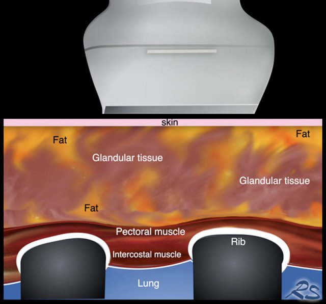
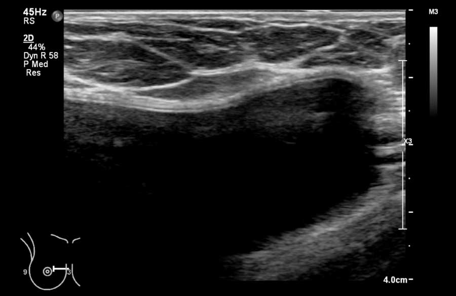
When we place a transducer on the breast, the first layer that we see is the skin with underneath a mixture of glandular and fatty tissue.
The deepest layer is the chest wall with the pectoral muscle, the ribs and the intercostal muscles.
Posterior or deeper to the ribs no imaging is possible due to the absorption of the sound waves and this results in an artefact called posterior acoustic shadowing.
This means that you cannot look beyond the ribs and that posterior to the ribs, the image is black.
The normal lungs are filled with air which also reflect ultrasound waves.
The anterior border of the lung produces a hyperechoic or white line which moves as a result of normal breathing (see video below).
The sound-reflection of the air-bubbles in the lungs produce what we call a dirty shadow (see next image).
This is not as pronounced as in the ribs.
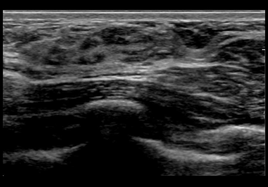
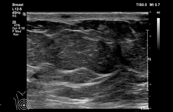
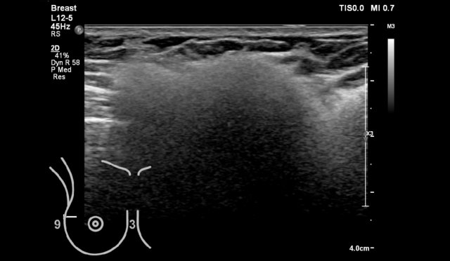
Here we see a normal ultrasound image of the breast.
The upper grey layer is the skin.
Then there is a mixture of fat (dark or hypoechoic) and glandular tissue (light grey or hyperechoic).
The striped layer posterior to the breast tissue is the pectoral muscle.
Posterior or deeper to the ribs there is a black area or posterior shadowing.
The lungs are the deepest visible layer.
The air in the lungs reflect most of the sound waves resulting in a bright or hyperechoic line with a dirty shadow posterior to it.
On this video normal breast anatomy.
Notice the movements of the lung during breathing.
In this video a breast cancer is seen within the glandular tissue.
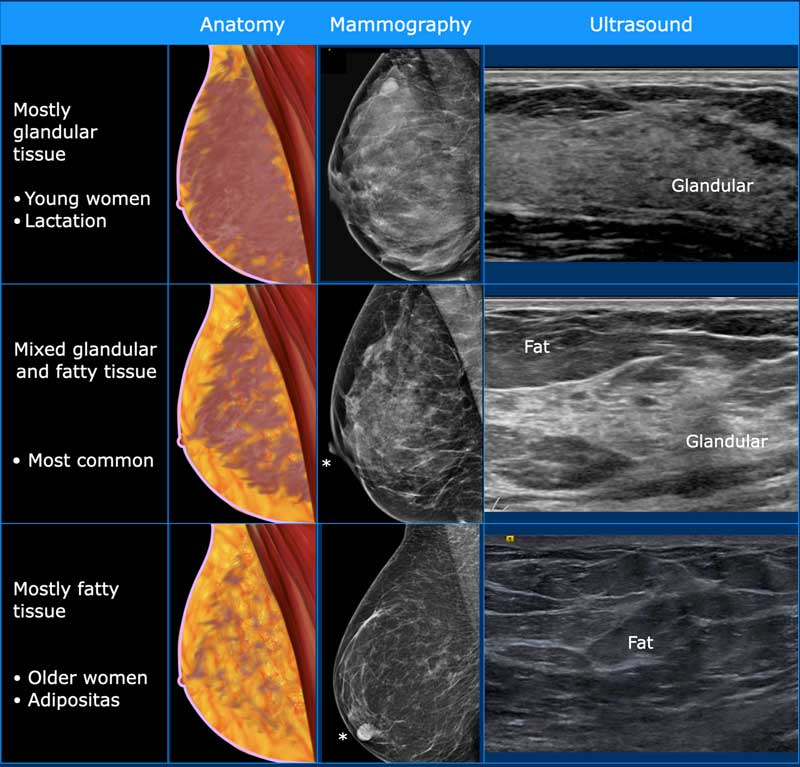
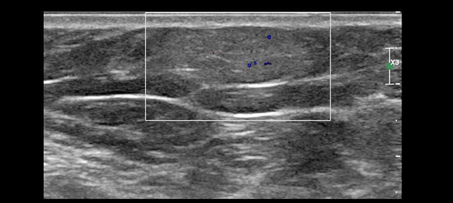
Breast composition
With ultrasound we can determine the composition of the breast: homogeneous fibroglandular - heterogeneous tissue - or homogeneous fat (fig).
Notice that the mammografic and ultrasound images are very much alike.
In young women the breast mostly contains glandular tissue.
This glandular tissue can be very pronounced during pregnancy and lactation and can show cyclic changes in premenopausal women resulting in breasts that feel lumpy or painful.
In older women the glandular tissue is gradually replaced by fat, although some older women still may have a reasonable amount of glandular tissue.
In adipose women there is more fat in the breasts even in younger women.

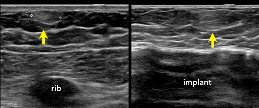
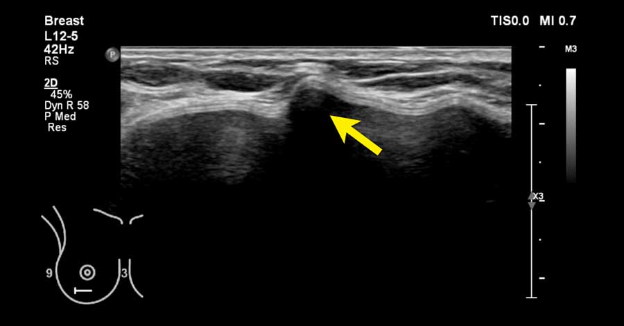
Near the midline the ribs are only composed of cartilage and are not calcified.
The cartilage does not produce a white echo on the anterior side or posterior shadowing.
Instead a hypoechoic structure is seen anterior the the lungs.
Do not mistake this structure for a breast tumor.
At first glance this may look like a fibroadenoma when you image the rib on cross section.
By turning the transducer you will notice that it is a long structure connected to the calcified part of the rib.
The small subcutaneous lesion is a dermoid cyst, which we will discuss later.
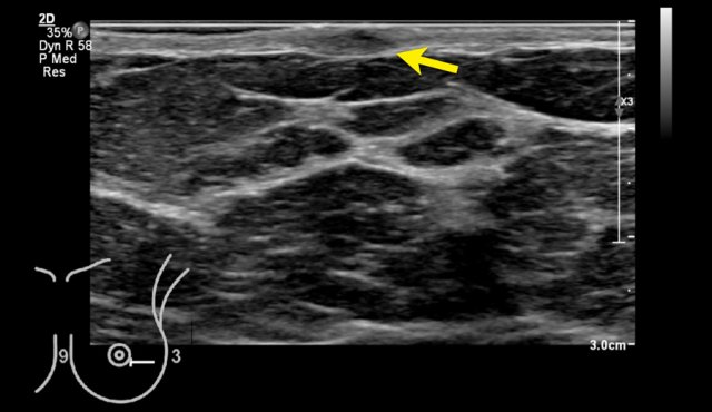
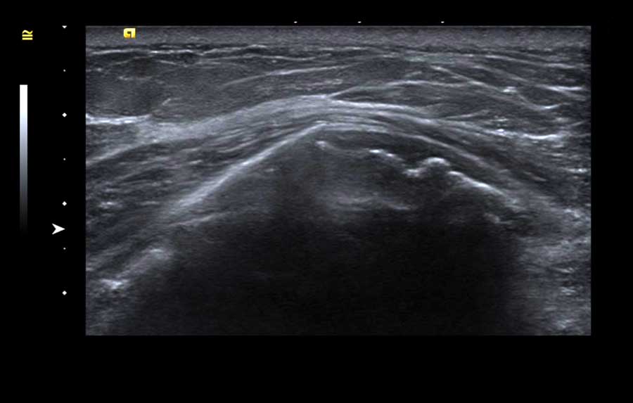
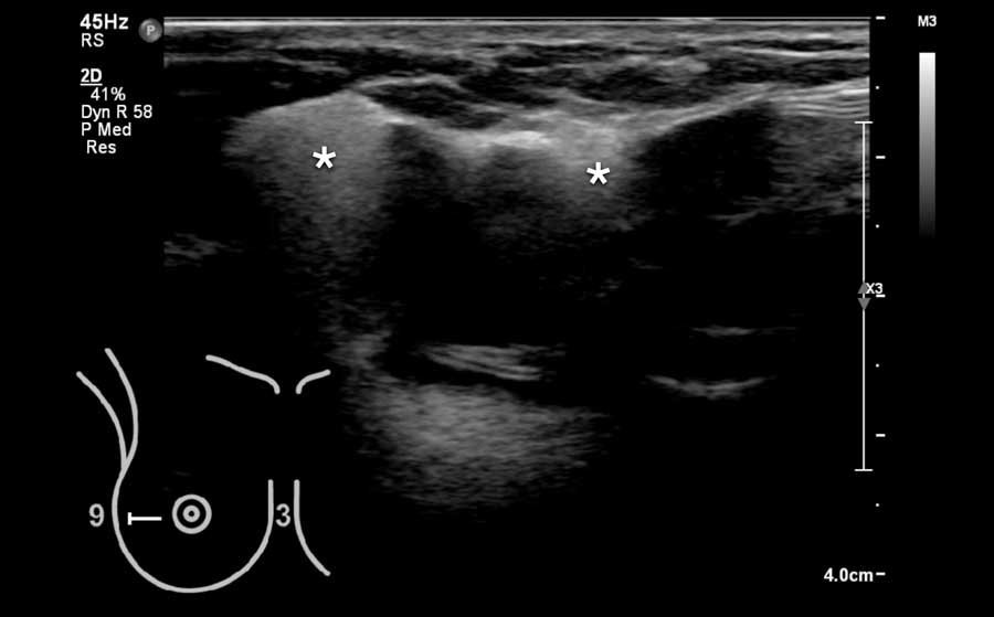
Within the same breast there may be areas with more fatty tissue and areas with mostly fibroglandular tissue as we can see on the video.
When you look at the contour of the glandular tissue (arrow), you can imagine, that this can feel bumpy on palpation and sometimes give the impression of a mass, when it is very pronounced.
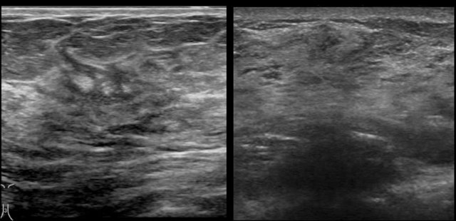
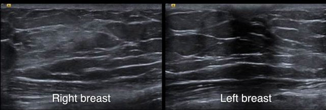
The way that the breasts present on an ultrasound image may differ between machines of different manufacturers.
This means that you have to get used to the images on your own ultrasound machine.
The images are of a Philips (left) and Siemens (right) ultrasound machine.
Look for instance at the difference in the presentation of the skin.
Ultrasound findings - overview
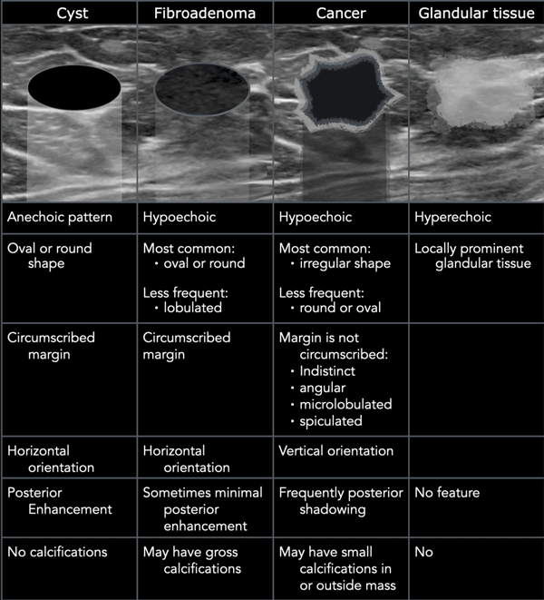
By far the most common abnormalities in the breast, which usually present as a lump in the breast are cysts, fibroadenomas, breast cancer and palpable glandular tissue.
In the table the typical ultrasound findings are listed (click to enlarge).
We will discuss each of these findings in more detail in a moment.
Cysts are seen in all age groups
Fibro-adenomas are benign tumors which are commonly seen in young women (especially 15-25 years) and seldom as a new finding in women over 50 years.
Breast-cancer is seen predominantly in women over 50 years of age and not that common in younger women.
Palpable glandular tissue is seen in young women, in pregnancy and breast-feeding.
It usually changes during the menstrual cycle.
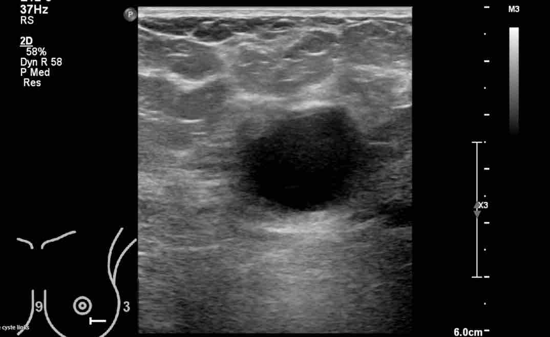
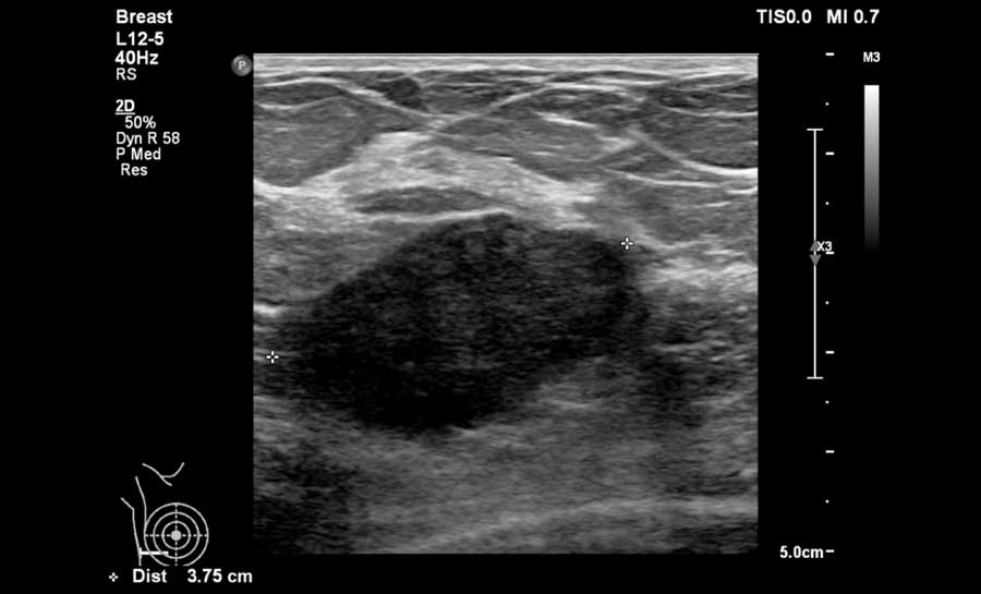
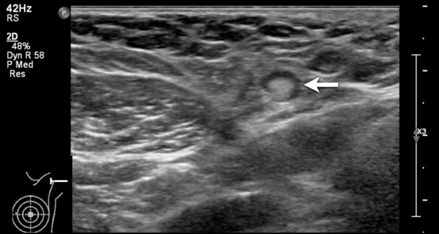
Cyst

Cysts are the most common lumps in the breast.
They are fluid-filled sacs inside the breast and are always benign.
It is extremely important to determine the cystic nature of a palpable lump, because then you can really reassure the patient that everything is fine.
On ultrasound the typical features of a cyst are:
- Echolucent (or black) pattern
- Round or oval shape
- Sharp circumscribed margin.
- Posterior enhancement
Posterior to a cyst, there is usually acoustic enhancement, also called posterior enhancement, which refers to the increased echoes deep to the cyst, because fluid transmits sound very well.
When the fluid is under tension, the cyst becomes more round and can be palpable.
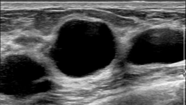
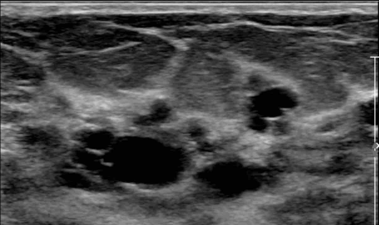
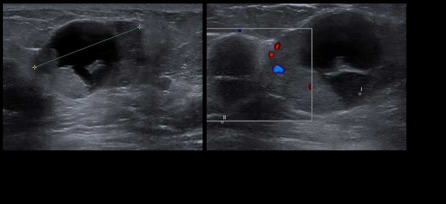
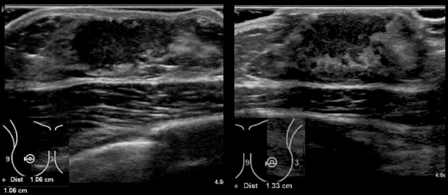
Here a typical example of multiple cysts in a woman who felt a lump in her breast.
Although there a many cysts, only the cyst in the center was palpable, because it was round with fluid under tension.
The other cysts were not palpable, because they just felt like the surrounding normal breast tissue.
It is very common to find more cysts in a woman who presents with a palpable cyst.
This woman had multiple small cysts in both breasts.
These cysts were not palpable.
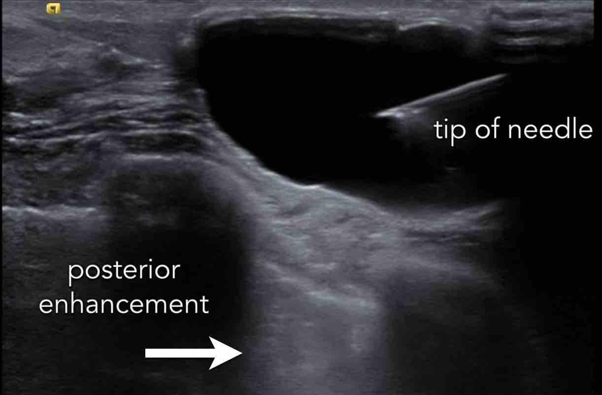
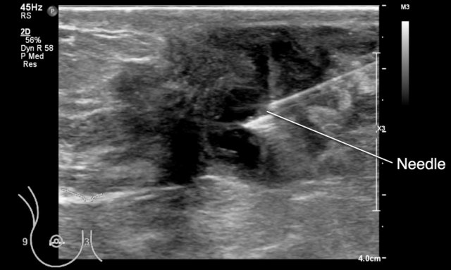
Here a video of a palpable cyst.
In this case the cyst was painful and it was decided to do a puncture with aspiration of the fluid.
Notice that the wall is a little bit thickened.
This is frequently a sign of low grade infection and explains why the cyst was painful.
Uncomplicated cysts are usually not painful.
This is another infected cyst, which was aspirated.
Aspiration is a quick and simple procedure.
In most cases the fluid has a transparent yellow color, but it can be green or brown.
Examination of the aspirated fluid is not necessary.
Complicated cyst
Most cysts have the typical appearance as shown above.
Complicated cyst have an atypical appearance:
- Hypoechoic content - the content of a cyst can become more proteinaceous and viscous due to infection or internal bleeding. As a result the echo pattern can become hypoechoic instead of the normal echolucent pattern.
Usually these complicated cysts still have posterior enhancement, what proofs the cystic nature of the lesion. - Thick wall - usually as a result of infection. Sometimes the wall can become irregular and then it can be difficult to differentiate these cysts from a cancer, especially if the content has become hypoechoic.
A complicated cyst with a hypoechoic content usually contains low-level echoes, that move when the patient changes in position.
When there is still doubt whether a lesion is a cyst or a solid tumor, then puncture with aspiration can solve this problem.
The image shows an echolucent, maybe somewhat hypoechoic lesion with some irregularity of the wall, which is thickened.
There is however posterior enhancement, which made us think, that this probably was a cyst.
A puncture was performed and the cyst was totally aspirated, which was the final proof.
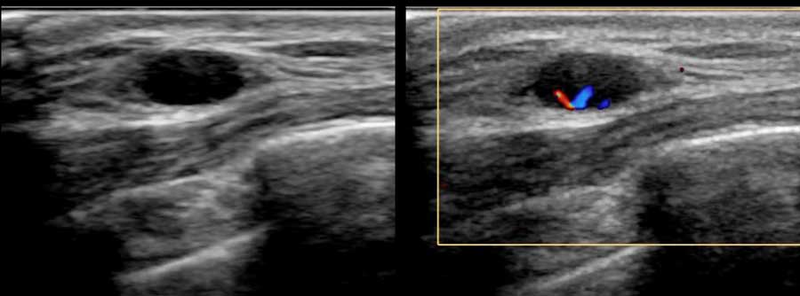
Intracystic tumor
Sometimes a part of a cyst is not echolucent, but hypoechoic or hyperechoic.
This can be the result of pus or debris, but it can also be an intracystic tumor like on these images.
Intracystic tumors are rare.
When you see vessels with color doppler then you know it is an intracystic tumor, which can be benign or malignant.
This is a tumor with a cystic component and not a cyst.
In absence of flow with color doppler and in absence of any low level echoes moving by changing patient position a puncture should be performed to differentiate between a complicated cyst and a solid mass.
Pus and debris can be aspirated unlike a tumor.
In this case biopsy revealed an intracystic carcinoma.
 Intracystic breast cancer
Here another breast cancer with a cystic component.
Notice the large solid component with flow on doppler image.
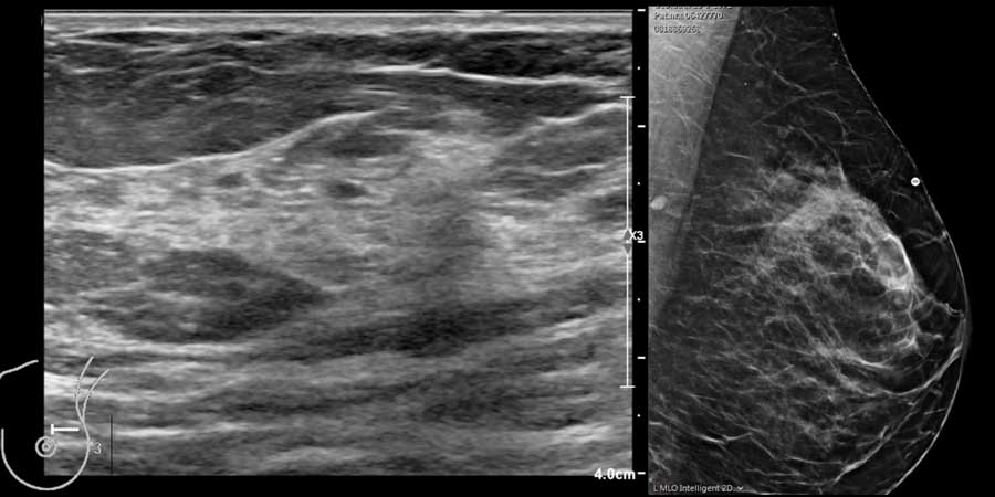
Fibroadenoma
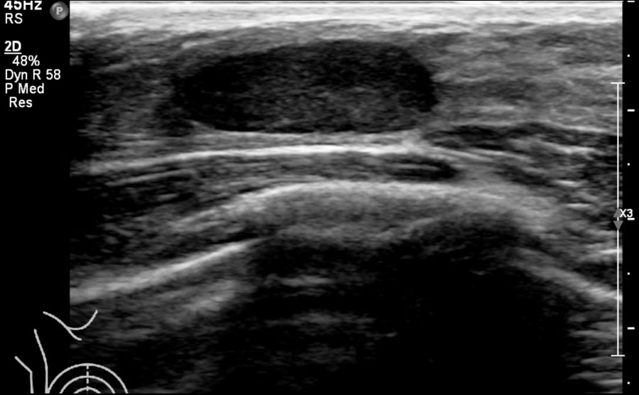
Fibroadenomas are benign tumors which are commonly seen in young women especially 15-25 years and seldom in women over 50 years.
The typical ultrasound features are:
- hypoechoic
- oval shape - sometimes round or with 2 or 3 lobulations.
- sharp border (also called circumscribed margin)
- orientation parallel to the skin (wider than tall)
- bloodflow within small vessels can sometimes be detected with color doppler
- variable a little bit of posterior enhancement
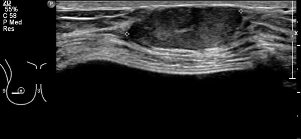
Here another typical fibroadenoma.
Notice that the margin is somewhat lobulated.
This woman has breast prostheses, which are seen as fluid-filled structures.
They were placed posteriorly to the pectoral muscle.
Scroll image for text.
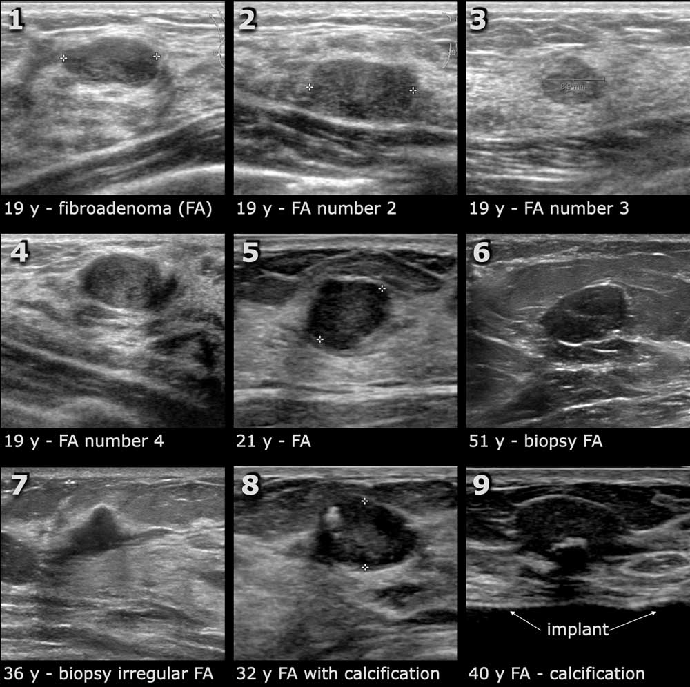
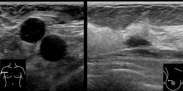
Here some examples of fibroadenomas.
Number 1-4 are in the same patient.
This is a common finding.
When you see one fibroadenoma, you can usually find more.
The lesion number 6 was biopsied because of the age of the patient and turned out to be a fibroadenoma.
Lesion number 7 was biopsied because it had an irregular shape and looked like a carcinoma.
This also turned out to be a fibroadenoma.
Fibroadenomas sometimes have calcifications but these are larger than the small calcifications that we encounter in carcinomas.
Microcalcifications as seen in ductal carcinoma in situ (DCIS), which can be a precursor of a carcinoma, are frequently not visible on ultrasound.
In the detection of DCIS mammography has advantages.
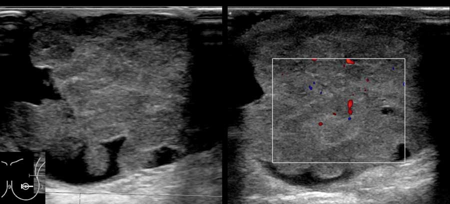
Breast cancer
Breast cancer is the most common malignant tumor in women.
A woman's risk of getting breast cancer increases with age.
Most women diagnosed with breast cancer are over the age of 50, but younger women can also get breast cancer.
The first noticeable clinical symptoms are:
- lump or area of thickened breast tissue
- retraction of the nipple
- dimpling of an area in the breast by retraction of underlying tumor
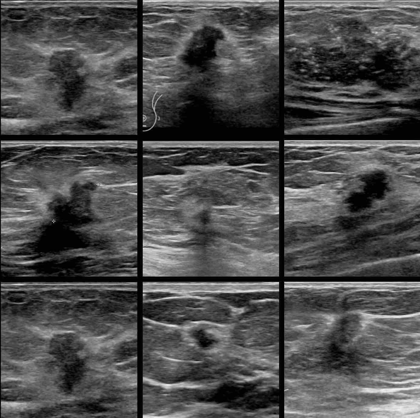
Here are some examples of breast cancer.
The key-features are:
- hypoechoic mass
- mostly irregular shape - sometimes round or oval and with orientation not parallel to skin
- indistinct border sometimes with halo
- may have posterior shadowing and small calcifications
We will now discuss the ultrasound findings in breast cancer in more detail.
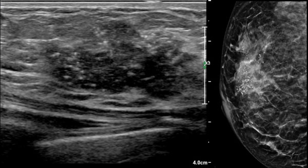
These images are of a 50-year-old woman who felt a lump in her breast.
Describe the ultrasound findings and then continue reading.
The ultrasound findings are:
- Hypoechoic mass with an irregular shape
- Uncircumscribed border which is both angular and indistinct
- Small hyperechoic dots, which represent calcifications
- No posterior shadowing or enhancement
These calcifications are also seen on the mammography.
The white area on the mammogram is the tumor.
Continue with the video.
Click on the image to start the video.
Click again on image to stop video.
Hyperechoic halo
A common finding in breast cancer is a hyperechoic halo surrounding the hypoechoic mass.
This halo is part of the tumor and should be included in the measurement of the tumor.
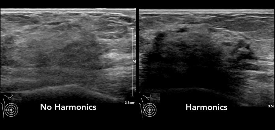
Harmonics
Harmonic imaging is an ultrasound technique that employs the resonance characteristics of tissue.
It is also calles tissue harmonic imaging or THI.
If you have this possibility on your ultrasound machine, you will notice that images produced with harmonic imaging have a higher resolution and are associated with fewer artifacts than conventional ultrasound imaging.
Posterior shadowing can be enhanced.
Notice that the small breast cancer is better seen with harmonic imaging.
There is a hypoechoic tumor with a hyperechoic halo and a little bit of posterior shadowing. The orientation is vertical.
The border is indistinct and the shape of the tumor is irregular.
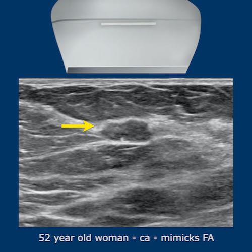
This is a difficult and uncommon case.
When you look at the image on the left a tumor is hardly seen.
It almost looks like normal glandular tissue.
However a mass was felt and when we look at the image with harmonics on, we can see the posterior shadowing.
This turned out to be a breast cancer.
If you have harmonics on your machine, it is best to view with and without harmonics in cases that aren't obvious to start with.
Examples of breast cancer
Scroll through eleven examples of biopsy proven breast cancers.
Video-examples of breast cancer
Click on the image to start the video.
Click again on image to stop video.
he video contains 3 examples of breast cancer.
Notice that the last video is of a 21-year-old woman with breastcancer.
This is uncommon, but unfortunately breast cancer is sometimes seen in young women.
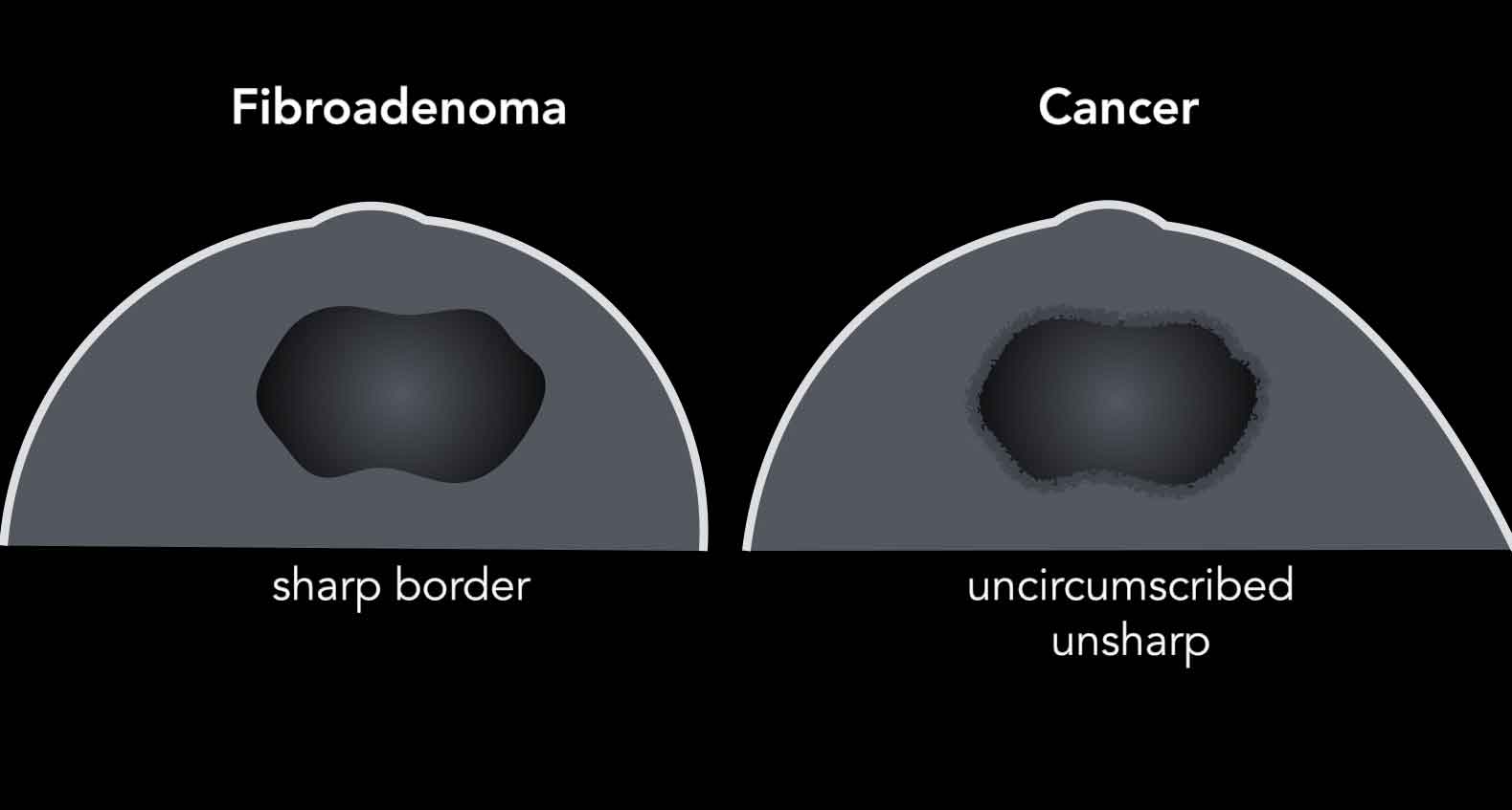
Breast cancer versus Fibroadenoma
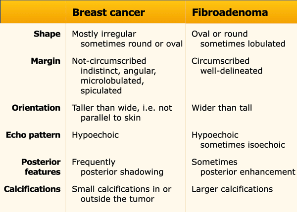
Sometimes breast cancer can look like a fibroadenoma and fibroadenomas can look like a cancer on ultrasound.
In the table the differences in ultrasound appearances are listed.
The age of the patient is another important issue, since fibroadenomas are commonly seen in young women especially 15-25 years and seldom in women over 50 years, while breast cancer is most commonly diagnosed in women over the age of 50 and is not common in younger women.
In some cases it is not possible to make the distinction by ultrasound imaging alone and a biopsy is needed for a final diagnosis.
Scroll through these images.
The differences between a fibroadenoma and a carcinoma are summarized.
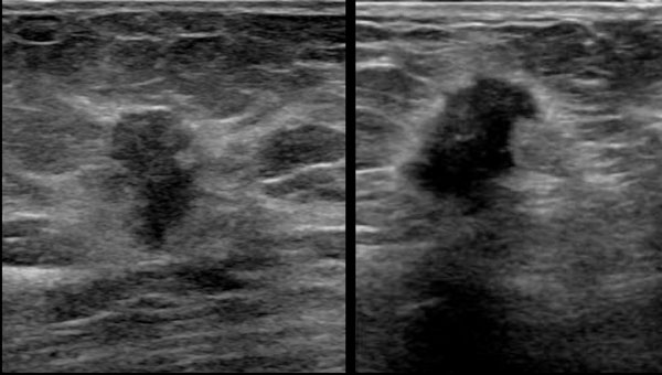
Here we have two oval-shaped hypoechoic lesions.
At first glance they look not quite different.
Study the images and determine the differences.
The lesion on the left is a carcinoma.
- Irregular shape
- Angular and indistinct border
- Hyperechoic halo
- Orientation is more vertical and not parallel to the skin, i.e. taller than wide
- Posterior shadowing
The lesion on the right is a fibroadenoma.
- Oval shape with slight lobulation
- Sharply demarcated
- Orientation is horizantal, parallel to the skin
- Posterior enhancement
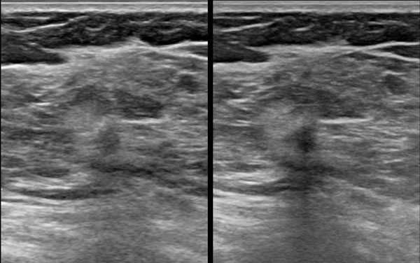
 Breast cancer: at first glance looks like a fibroadenoma
Age
How does the age of a woman help us in the differentiation between a fibroadenoma and breast cancer?
In most cases it confirms our imaging diagnosis.
On the other hand when we see a mass in the breast in a younger woman that does not full-fill all the criteria of a typical fibroadenoma, we should do a biopsy to rule out a carcinoma.
For instance because the lesion is not circumscribed or taller than wide.
The same holds true for a new mass that looks like a fibroadenoma in an older woman.
The image shows a lesion, that at first glance looks like a fibroadenoma.
Yet there are two things that don't fit.
First the age of the woman is 49 years and secondly on the posterior side the contour shows some irregularities.
A biopsy was performed and the mass proved to be a cancer.
Palpable glandular tissue
Fibroglandular tissue is not evenly spread in the breast and is usually more pronounced in the upper lateral quadrant.
Sometimes focal swelling of glandular tissue can be very pronounced and causes a lump, that on palpation cannot be differentiated from a tumor.
The ultrasound shows a collection of glandular tissue within a fatty breast.
Since the glandular tissue is more firm than the fatty tissue, this feels like a mass on palpation.
A mammogram was performed with a marker on the palpable mass and also showed a focal collection of normal glandular tissue.
The video is of a woman who felt a lump in her breast.
On ultrasound pronounced glandular tissue was found.
Since the glandular tissue is more firm than the surrounding fatty tissue, you can imagine that when you glide with your finger over the skin, this area would feel like a bump.
Fibroglandular tissue can be so pronounced that it is difficult for the ultrasound beam to pass through the tissue.
This may give the impression of an irregular hypoechoic mass with posterior shadowing and simulate a carcinoma (video).
However when you compress the tissue, you will see that it is just hyperechoic pronounced fibroglandular tissue.
Usually these are young patients who present with a large painful lump in their breast.
Abscess
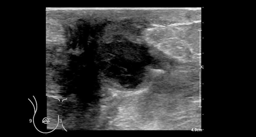
A breast abscess is a painful build-up of pus in the breast caused by an infection.
It mainly affects women who are breastfeeding as a complication of mastitis and is uncommon in non-breast feeding women.
Women usually present with a painful lump and the skin can be red and thickened.
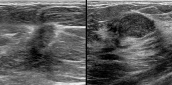
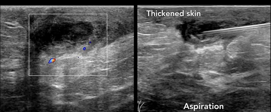
The ultrasound is of a woman who presented with fever and a painful lump in the breast behind the retracted nipple.
During mild compression and decompression with the transducer it was noted that the fluid in the abscess was moving around.
Notice also the posterior enhancement which is another indication that the structure contains fluid.
Continue with next image...
Subsequently the abscess was apirated.
Aspiration is the first choice treatment of abscesses.
No need for surgery or antibiotics in this case, although antibiotics are sometimes also given.
Here another abscess located behind the nipple, which was aspirated.
This can be a painful procedure.
It is best to inject local anesthesia in the skin and subcutis and to try to drain the pus through that same needle.
If the pus is too thick, a thicker needle will do the job.
Take some time for the local anesthesia to work.
Skin and subcutis
Abnormalities that originate in the skin or subcutis may present as a lump in the breast, but they do not represent breast abnormalities.
When you can determine that a lesion originates in the skin or subcutis, you know that you are not dealing with a breast tumor.
The ultrasound images show a lesion that is located in the skin and not within the breast.
Most of these lesions are dermoid cysts.
Try to find the connection to the skin, although this is not always visible.

This woman presented with a painful lump in her breast and the skin was red.
This is another intradermal lesion.
Notice that the skin is thickened.
This probably is a dermoid cyst with inflammation.
It healed without any treatment.
Here another dermoid cyst.
It is located in the subcutis and connected to the skin.
This is not a breast tumor.
Scroll through more benign lesions that originate in the skin or subcutis.
Lipoma
Lipomas are not that common in the breast, but when they occur, they just look like any other lipoma in the body.
They are iso- or hyperechoic compared to the surrounding fat.
They are always benign.
Sometimes a lipoma cannot be differentiated from fat necrosis, which we will discuss now.
Fat necrosis
Fat necrosis is a benign entity frequently presenting as a superficially located palpable small mass within the breast.
It is thought that fat necrosis is the result of trauma although the patient often does not recall a specific traumatic event.
As a result of the trauma, the fatty tissue undergoes changes which make it more firm and sometimes tender on palpation.
On ultrasound it usually presents as a part of the fatty tissue that is mildly swollen and hyperechoic compared to the surrounding fatty tissue.
Hence the similarity to a lipoma.
The content may liquify and result in an oil cyst, which on ultrasound just looks like any other cyst.
A lipoma is usually more soft and non-tender.
Ribs
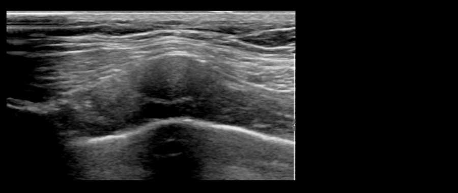
A protruding rib can cause a hard swelling which may simulate a breast tumor.
This patient complained of a painful hard swelling within her breast.
On ultrasound a protruding costochondral junction was seen.
This is the part where the calcified part of the rib meets the cartilage part.
Frequently calcifications can be seen within the cartilage like in this case.
This video shows normal rib cartilage.
By turning the transducer you can see that the rib is a longitudinal structure anterior to the lung and posterior to the pectoral muscle..
Axilla
Normal axillary lymph nodes are usually small oval shaped hypoechoic structures with a hyperechoic centre.
The hyperechoic centre is mainly composed of fat and contains the vessels.
The surrounding hypoechoic cortex is the actual lymphogenic tissue.
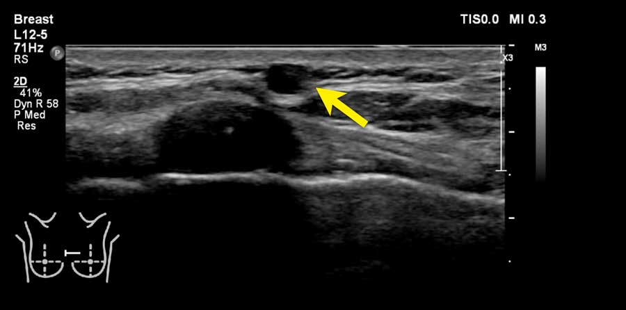
 A normal intramammary lymph node
Sometimes a lymph node only presents as an oval hypoechoic lesion without a visible hyperechoic center.
When you see vessels in the hilus like in this case, then it definitely is a lymph node.
In fact this lymph node was located within the breast, i.e. an intramammary lymph node, which is a benign finding.
 Large axillary lymph node metastases of a small irregular carcinoma
Enlarged lymph nodes
Enlarged lymph nodes in the axilla can be the result of lymphogenic metastatic disease of breast cancer.
The images are of a 50-year-old woman who presented with palpable masses in the axilla, that represented round enlarged hypoechoic lymph nodes.
Subsequently there was an ultrasound examination of the breast, which revealed a small non-palpable hypoechoic irregular cancer.
Ectopic glandular tissue in the axilla
Some women have fibroglandular breast tissue in the axilla.
This tissue behaves just like the glandular tissue in the breast and may get tense or painful at some time in the menstrual cycle.
The ultrasound image is of a young woman who felt a painful swelling in her axilla.
The image shows normal glandular tissue in an atypical location.
Breast implants
Most breast implants are silicone-filled prostheses.
On ultrasound they look like a large cyst.
The implant is most commonly placed posterior to the breast tissue or posterior to the pectoral muscle.
Implants filled with silicone or saline are anechoic (echolucent).
In most patients, breast implants have a regular aspect with lobulated margins.
Sometimes this lobulation can give the impression of a lump in the breast (figure).
 Collections or silicone leakage **
Silicone leakage
When silicone leaks out of the implant it will cause a hyperechoic shadow or dirty shadow, just like we can see in the lungs caused by the air in the lungs.
The image shows leakage of silicone anterior to the prosthesis.
Free silicone breast injections are an alternative form of breast augmentation to breast implants.
They have serious adverse effects and are banned in many countries.
The whole image looks blurred.
This is called snow storm appearance.
You will get the same image as when there is massive leakage.
Here a fibroadenoma anterior to a silicone implant.
Ultrasound in Men
Gynecomastia
Gynecomastia is the most common male breast disorder and commonly presents as a palpable lump or tenderness of the breast.
Clinically, gynecomastia presents as a soft mobile tender subareolar mass.
Any mass that is not subareolar is not gynecomastia.
On ultrasound the abnormality should be located right behind the nipple.
Breast cancer in men just looks like breast cancer in women.
These images are of 70-year-old male who presented with a painfull swelling behind the right nipple.
Notice that there is some fibroglandular tissue on the right, while on the left there is only subcutaneous fat.
 Bilateral gynecomastia in an athlete taking steroids
Newborns, boys going through puberty and older men may develop gynecomastia as a result of normal changes in hormone levels.
A common cause of gynecomastia is the use of anabolic steroids used by athletes to build muscle and enhance performance.
This patient had used steroids and has gynecomastia on both sides.
Sometimes gynecomastia can result in images that simulate a carcinoma, like on the images here, but luckily the findings were on both sides and there was a history of steroid intake.
The diagnosis of gynaecomastia is usually easier to make on a mammogram.
Nipple region

The region of the nipple can be difficult to examine.
In most cases you see only some thickening of the skin and behind the nipple some dilated ducts.
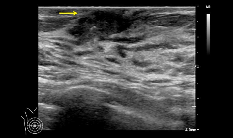
This image is of a patient who presented with a retracted nipple.
There is an irregular tumor behind the nipple with ingrowth into the nipple (arrow).
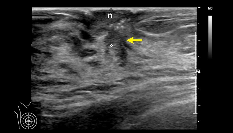
Here another tumor behind the nipple (n).
These tumors can be very difficult to detect.
In every woman who complains of a retracted nipple this area should be examined carefully.
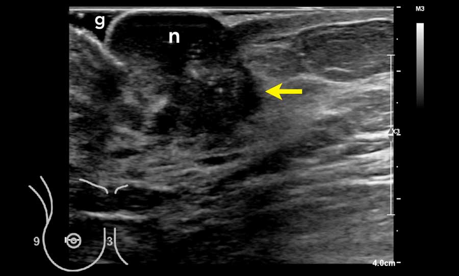
This patient had a large nipple, which made it difficult to examine the region behind the nipple.
A lot of gel (g) was used to get a good contact with the skin.
A large tumor is seen behind the nipple.