Cervical Lymph Node Map
modified from Robbins
Aurelia Fairise and Robin Smithuis
Institut de Cancérologie de Lorraine in Nancy, France and the Alrijne hospital in Leiderdorp, the Netherlands
Publicationdate
This article is based on the nomenclature proposed by the American Head and Neck Society and the American Academy of Otolaryngology-Head and Neck Surgery.
10 node groups are defined with a concise description of their main anatomic boundaries, the normal structures juxtaposed to these nodes, and the main tumor sites at risk for harboring metastases in those levels [1].
Overview
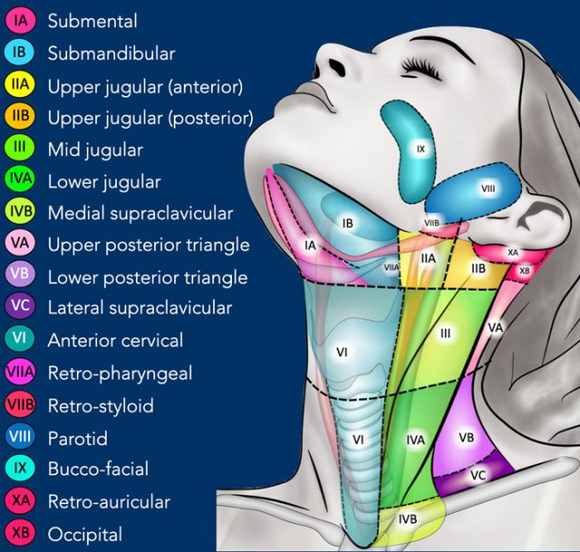
In this cervical lymph node map the levels were extended to 10.
Some of these are being divided into sub-levels to correspond more completely with the TNM atlas.
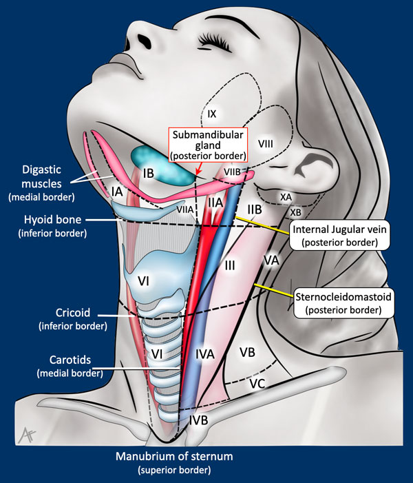
Borders
Important landmarks are:
- Hyoid bone
- Cricoid
- Carotids
- Sternocleidomastoid muscle
- Manubrium of sternum
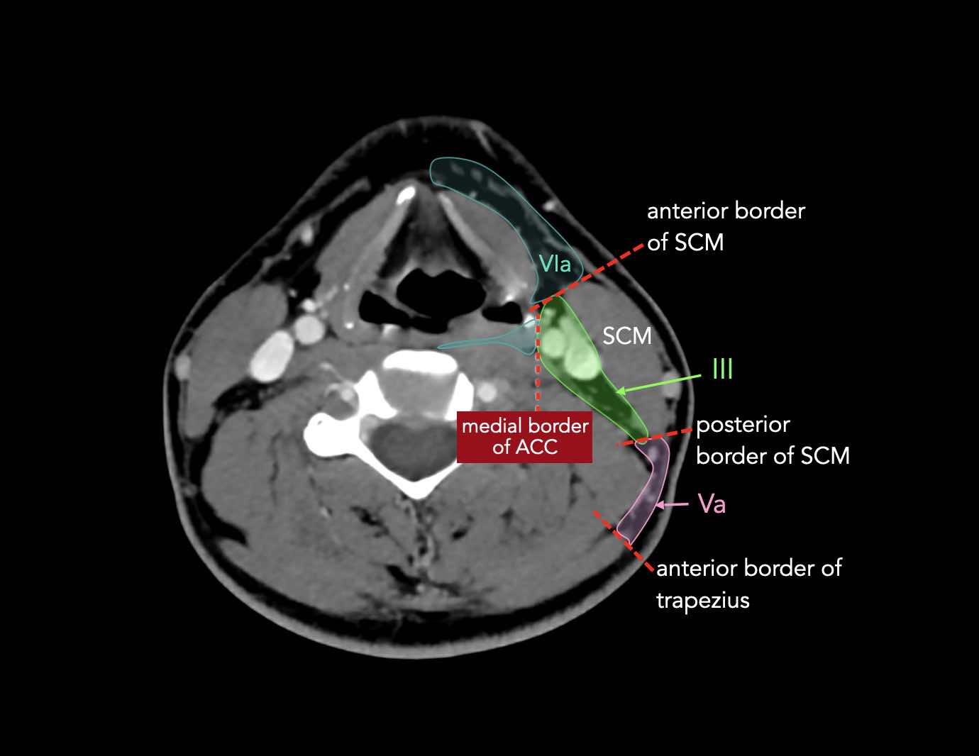
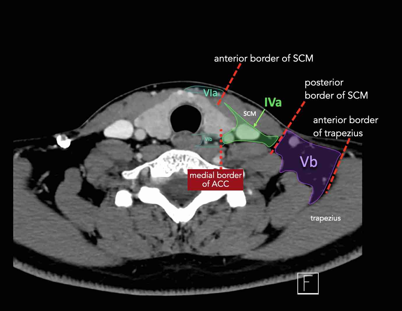
Axial CT
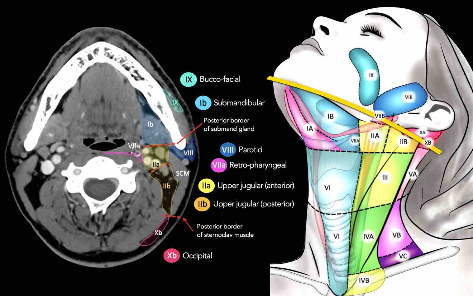
Axial CT slices in correlation to overview illustration.
Axial CT slices in more detail.
Enlarge images by clicking on them.
Levels
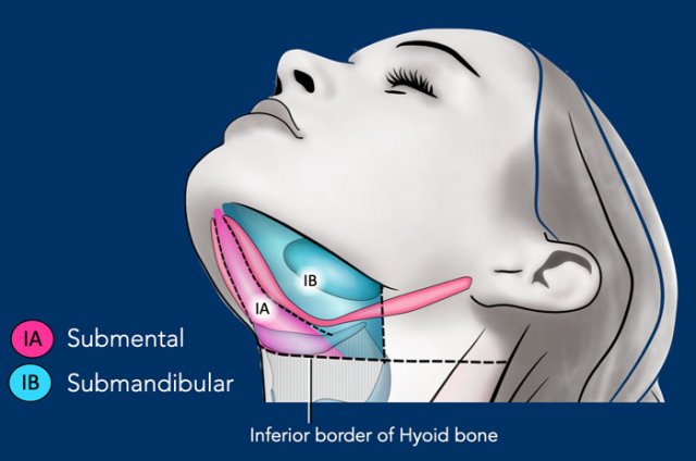
I - Submental and submandibular
Nodes in level I are at risk of developing metastases from cancers of the oral cavity, anterior nasal cavity and the soft tissues of the mid-face and the submandibular gland.
Level Ia
is a median region located between the anterior belly of the digastric muscles, which contains the submental nodes.
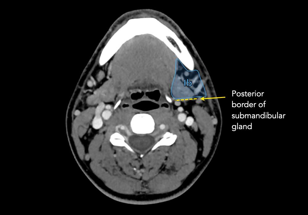
Level Ib
contains the submandibular nodes located in the space between the inner side of the mandible laterally and the digastric muscle medially, from the symphysis menti anteriorly to the submandibular gland posteriorly.
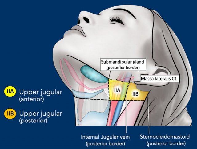
II - Upper jugular
Level II receives lymphatics from the face, the parotid gland, and the submandibular, submental and retropharyngeal nodes.
Level II also directly receives the collecting lymphatics from the nasal cavity, the pharynx, the larynx, the external auditory canal, the middle ear, and the sublingual and submandibular glands [1].
Level II can be divided into level IIa and level IIb by drawing a line at the posterior edge of the internal jugular vein.
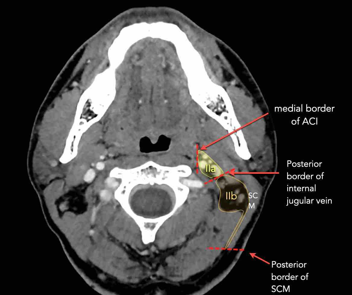
The nodes in level IIa and IIb are at risk of harboring metastases from cancers of the nasal and oral cavity, nasopharynx, oropharynx, hypopharynx, larynx and major salivary glands.
Level IIb is more likely associated with primary tumors of the oropharynx or nasopharynx, and less frequently with tumors of the oral cavity, larynx or hypopharynx [1].
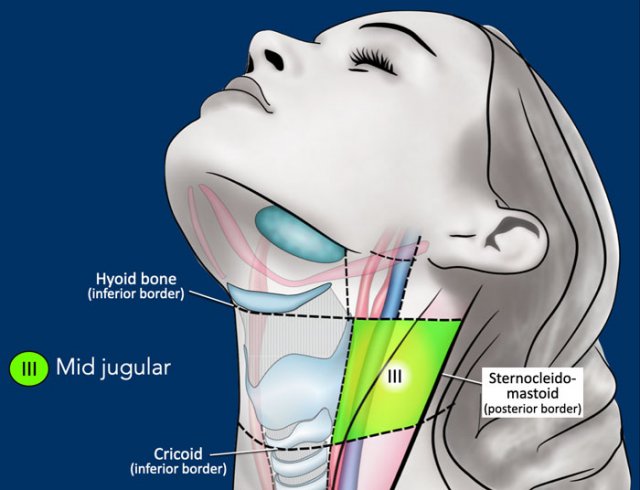
III - Mid jugular
Level III receives efferent lymphatics from levels II and V, and some efferent lymphatics from the retropharyngeal, pretracheal and recurrent laryngeal nodes.
It collects the lymphatics from the base of the tongue, tonsils, larynx, hypopharynx and thyroid gland.
The inferior border of the cricoid is the border between level III and IVA.
Nodes in level III are at risk of harboring metastases from cancers of the oral cavity, nasopharynx, oropharynx, hypopharynx and larynx.
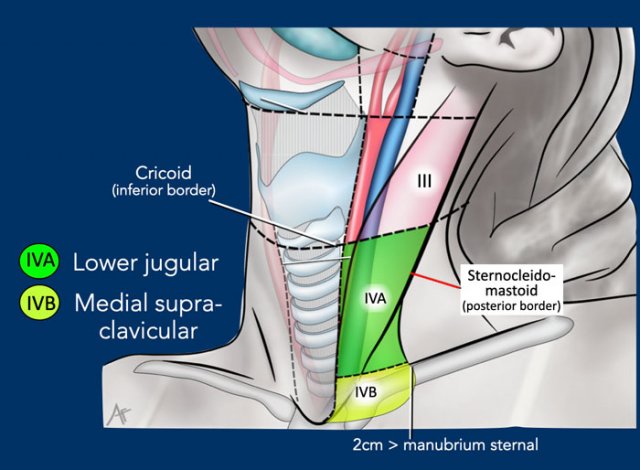
IV - Lower jugular and medial supraclavicular
The border between level IVa and IVb is set arbitrarily 2 cm cranial to the sterno-clavicular joint.
Level IVa
These nodes are at risk for harboring metastases from cancers of the hypopharynx, larynx, thyroid and cervical esophagus.
Rarely metastases from the anterior oral cavity may manifest in this location with minimal or no proximal nodal disease.
Level IVb
These nodes are at risk for harboring metastases from cancers of the hypopharynx, subglottic larynx, trachea, thyroid and cervical esophagus.
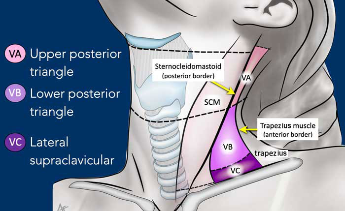
V - Posterior triangle and Supraclavicular
Level V contains the nodes of the posterior triangle group located posteriorly to the sternocleidomastoid muscle around the lower part of the spinal accessory nerve and the transverse cervical vessels.
Nodes in level V are most often associated with primary cancers of the nasopharynx, the oropharynx, the cutaneous structures of the posterior scalp, and the thyroid gland.
Level Vc - Supraclavicular
This level contains the lateral supraclavicular nodes located in the continuation of the posterior triangle nodes (level Va and Vb) from the cervical transverse vessels down to a limit set arbitrarily 2 cm cranial to the sternal manubrium.
It corresponds partly to the area known as the supraclavicular fossa.
Level Vc receives efferent lymphatics from the posterior triangle nodes (level Va and Vb) and is more commonly associated with nasopharyngeal tumors [1].
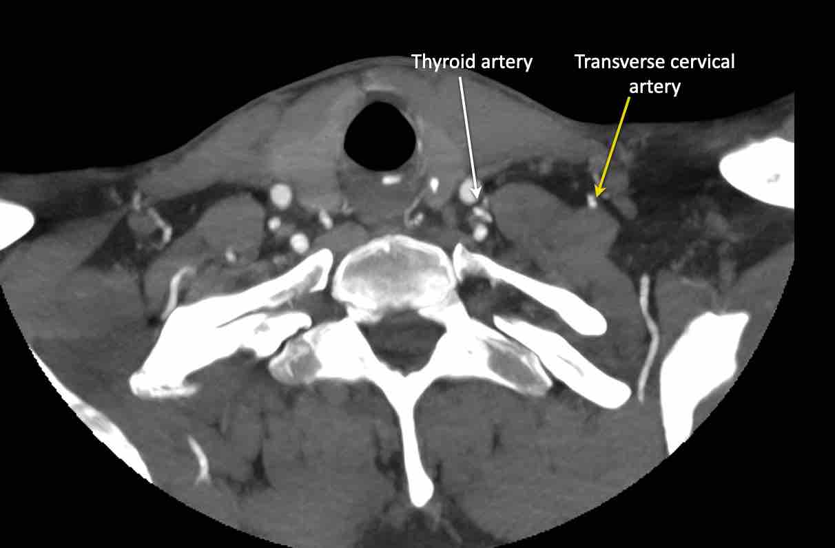
Transverse cervical artery
Scroll through the images for the anatomy of the transverse cervical artery.
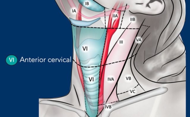
VI - Anterior cervical
This level contains the superficial anterior jugular nodes (level VIa) and the deeper prelaryngeal, pretracheal, paratracheal and recurrent laryngeal nerve nodes (level VIb).
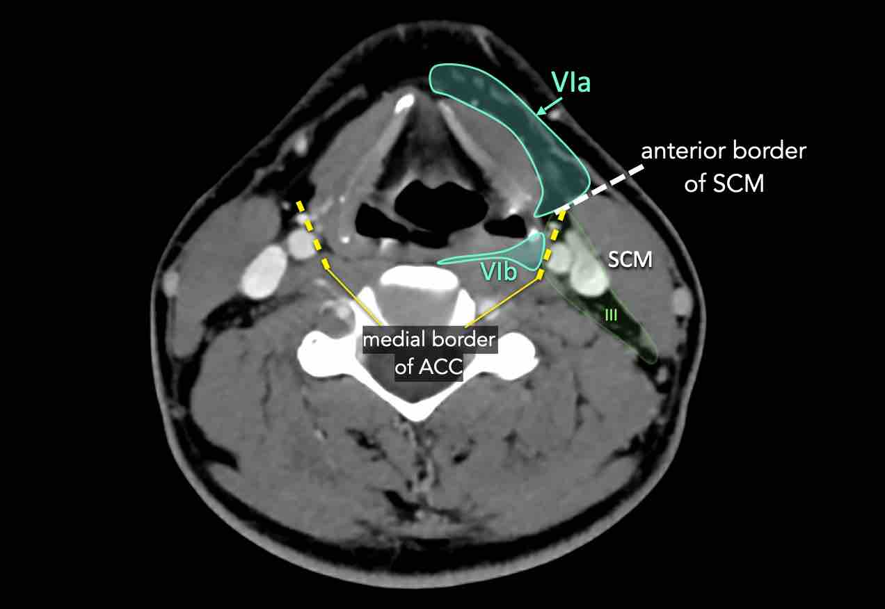
Level VIa
This level contains the superficially located anterior jugular nodes.
Level VIb
This level is contained between the medial borders of the common carotid arteries.
The nodes in this area are:
- pre-laryngeal nodes in front of the larynx and cricoid
- pre-tracheal nodes in front of the trachea
- paratracheal nodes also called recurrent laryngeal nerve nodes

Delphian lymph node
The Delphian lymph node derived its name from the oracle of Delphi, whose prophecy would be a death secondary to laryngeal cancer.
It is a pretracheal node in level VIa located anterior to the cricoid and in between the cricothyroid muscles.
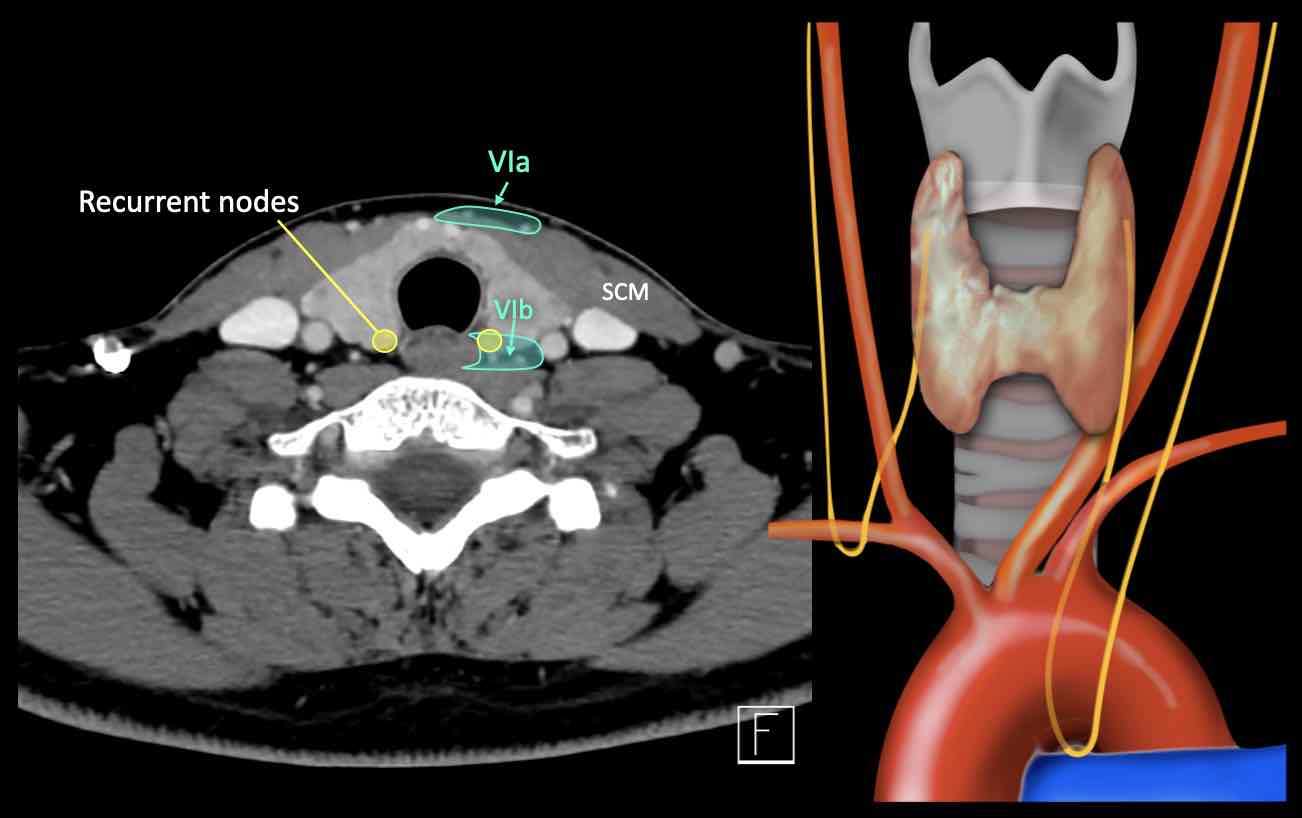
The recurrent laryngeal nerves branch off the vagus, the left at the aortic arch, and the right at the right subclavian artery.
The left laryngeal nerve can be compressed by subaortic lymph node metastases in the aorto-pulmonary window as seen in patients with lung cancer.
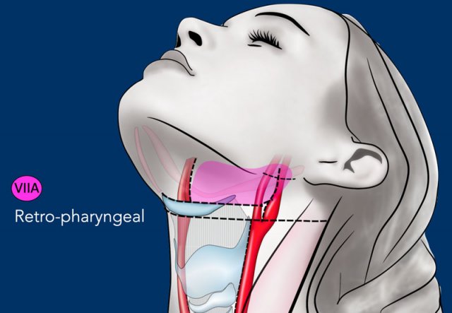
VII - Retropharyngeal and retrostyloid
Retropharyngeal nodes receive lymphatics from the mucosa of the nasopharynx, the Eustachian tube and the soft palate.
These nodes are at risk of harboring metastases from cancers of the nasopharynx, the posterior pharyngeal wall and the oropharynx (mainly the tonsillar fossa and the soft palate).
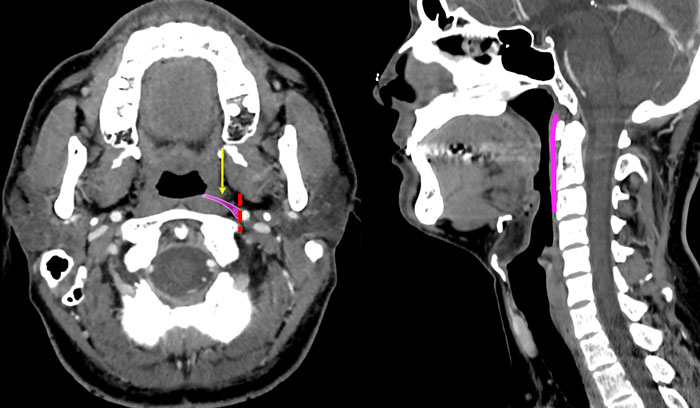
Level VIIa - retropharyngeal
These nodes lie within the retropharyngeal space, extending cranially from the upper edge of the first cervical vertebrae (massa lateralis) to the cranial edge of the body of the hyoid bone caudally (figure).
This space is bounded anteriorly by the pharyngeal constrictor muscles and posteriorly by the longus capitis and longus colli muscles.
Laterally, the retropharyngeal nodes are limited by the medial edge of the internal carotid artery.
Retropharyngeal nodes receive efferent lymphatics from the mucosa of the nasopharynx, the Eustachian tube and the soft palate.
These nodes are at risk of harboring metastases from cancers of the nasopharynx, the posterior pharyngeal wall and the oropharynx (mainly the tonsillar fossa and the soft palate).
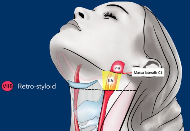
Level VIIb - retrostyloid
The retro-styloid nodes are the cranial continuation of the level II nodes.
They are located in the fatty space around the jugulo-carotid vessels up to the base of skull at the jugular foramen.
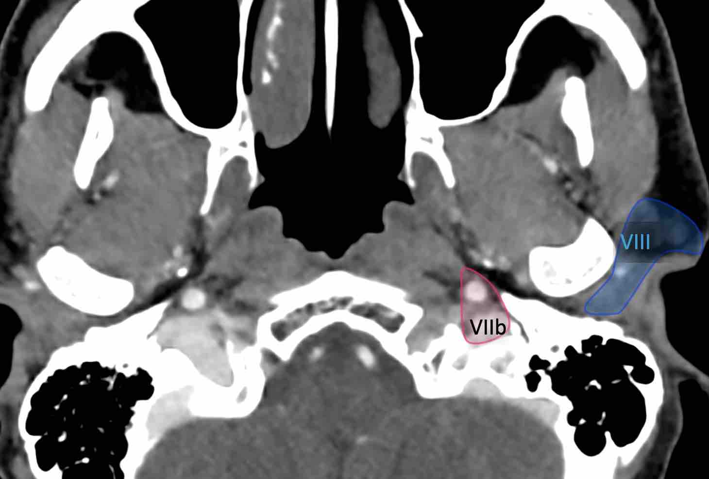
The retro-styloid space is delineated by the internal carotid artery medially, by the styloid process and the deep parotid lobe laterally, by the vertebral body of C1 and the base of skull posteriorly and by the pre-styloid para-pharyngeal space anteriorly.
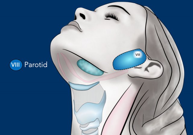
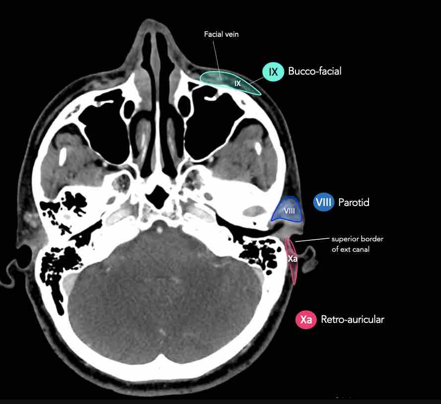
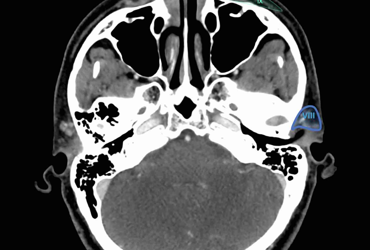
VIII - Parotid
This level contains the parotid node group, which includes the subcutaneous pre-auricular nodes, the superficial and deep intraparotid nodes and the subparotid nodes.
These nodes extend from the zygomatic arch and the external auditory canal down to the mandible.
They extend from the subcutaneous tissue laterally to the styloid process medially, and from the posterior edge of the masseter and the pterygoid muscles anteriorly to the anterior edge of the sternocleidomastoid muscle and the posterior belly of the digastric muscle posteriorly [1].
The parotid group receive lymphatic from the frontal and temporal skin, the eyelids, the conjunctiva, the auricle, the external acoustic meatus, the tympanum, the nasal cavities, the root of the nose, the nasopharynx, and the Eustachian tube.
They are at risk of harboring metastases from cancers of the frontal and temporal skin, orbit, external auditory canal, nasal cavities and parotid gland.
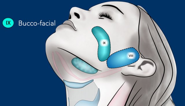
IX - Buccofacial
Level IX contains the malar and bucco-facial node group, which includes inconsistent superficial lymph nodes around the facial vessels on the external surface of the buccinator muscle.
These nodes extend from the caudal edge of the orbit (cranially) down to the caudal edge of the mandible (caudally) where they reached level Ib.
They lay on the buccinators muscle (medially) in the sub-cutaneous tissue, from the anterior edge of the masseter muscle and the Bichat’s fat pad (posteriorly) to the anterior sub-cutaneous tissue of the face.
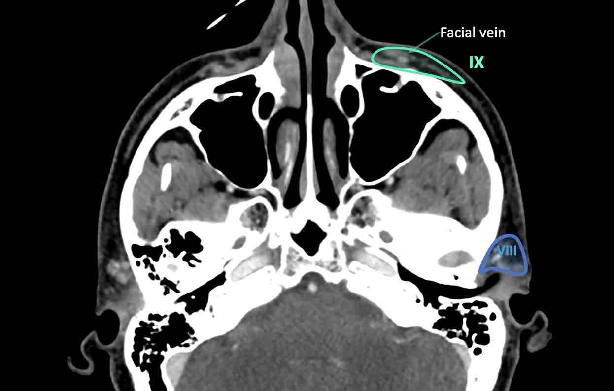
The bucco-facial nodes receive efferent vessels from the nose, the eyelids, and the cheek.
They are at risk of harboring metastases from cancers of the skin of the face, the nose, the maxillary sinus (infiltrating the soft tissue of the cheek) and the buccal mucosa.
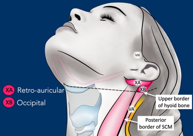
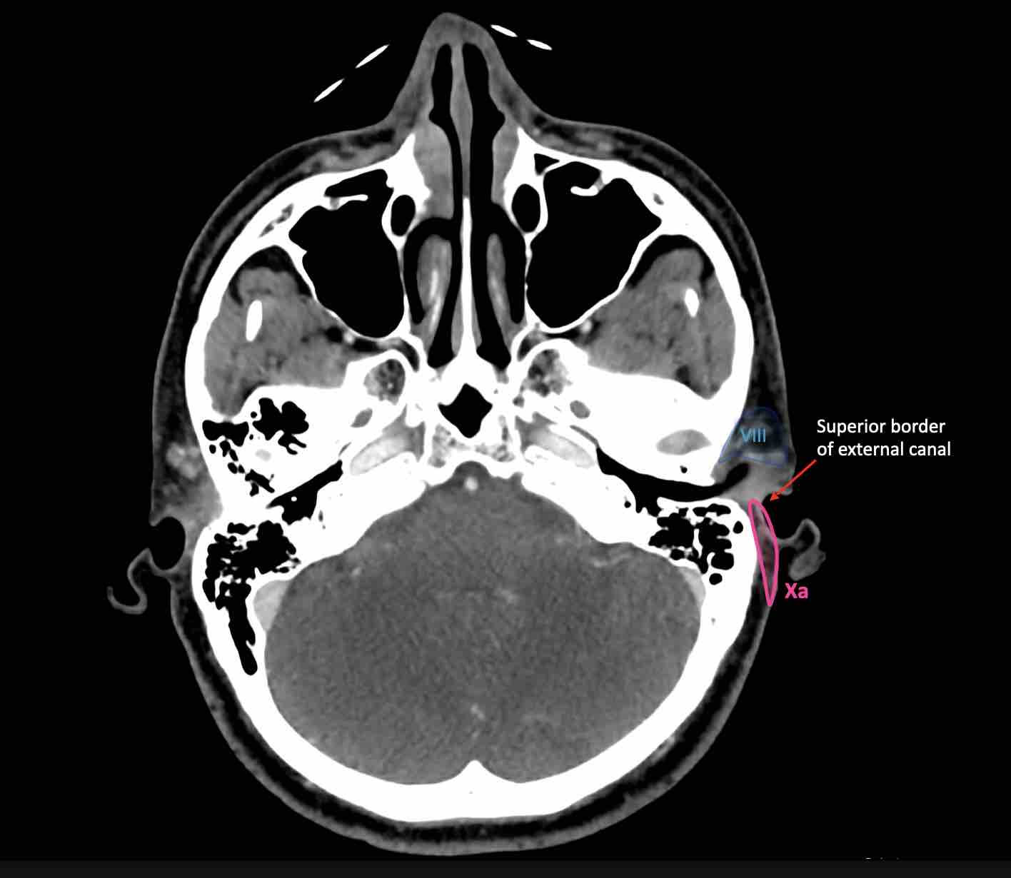
X - Retroauricular and occipital
Level Xa contains the retroauricular (also called mastoid) and subauricular nodes, which includes superficial nodes lying on the mastoid process from the cranial edge of the external auditory canal cranially to the tip of the mastoid caudally.
Level Xb contains the occipital lymph nodes, which are the cranial and superficial continuation of the level Va nodes up to the cranial protuberance. They lie from the posterior edge of the sternocleidomastoid muscle to the anterior (lateral) edge of the trapezius muscle.
Lymph node metastases in level X are from skin cancers of the retro-auricular area (Xa) and skin cancers of the occipital area (Xb).
Click on the image below to get more information about Medical Action Myanmar, a medical organization run by Nini Tun and Frank Smithuis, who happens to be the brother of Robin Smithuis.