BI-RADS v2025 Manual - Ultrasound
Updated version
Laura Schijf¹ and Robin Smithuis²
¹Radiology department of University Medical Center Amsterdam and ²Alrijne Hospital, Leiden, the Netherlands
Publicationdate
This article is an overview of the updated BI-RADS® Manual v2025.
We encourage all breast imaging professionals to consult the illustrated BI-RADS® Manual for comprehensive understanding and correct application of the system.
The BI-RADS v2025 Manual on Ultrasound builds on prior editions, reinforcing ultrasound’s indispensable role in breast imaging.
Since 2013, breast density reporting laws have been widely enacted, requiring patient notification of breast density. This has increased the use of supplemental ultrasound screening.
Major Updates:
- Breast Composition Assessment
- Lobulated shape
- Mixed solid and cystic mass
- Non-Mass Lesions
- Echogenic rind
- Lymph Node Evaluation
Ultrasound Lexicon

Key Updates in BI-RADS v2025 for Ultrasound
- Assessment of Breast Composition.
Describe the tissue pattern and the glandular component.
Studies reveal that ultrasound assessment of breast tissues may influence detection and indicate risk. This is significant in practices where ultrasound is used as a screening modality (or even the sole screening modality). - Lexicon Refinements
- Masses - Shape - Lobulated: Added to oval, round, and irregular for more precise communication.
- Masses - Echo pattern -Mixed solid and cystic: Emphasizes the importance of the solid component.
- Associated features - Echogenic rind: Describes the reaction in surrounding tissues that may accompany some cancers, invoking a sense of importance.
- New Finding Category
- Non-mass lesions. These are discrete findings (not simply heterogeneous breast tissue).This category aims to:
Expand ultrasound utility in diagnosing infiltrative pathologies, including lobular and in-situ breast cancers. - Expanded Discussion on Lymph Nodes.
- Nearly all breast cancers spread via the lymphatic system, making lymph node evaluation critical.
- Ultrasound is a key modality for node assessment, especially for biopsy guidance.
- The manual now includes: morphological changes in nodes involved by tumors and anatomy of regional nodal basins for TNM staging.
Breast compositon
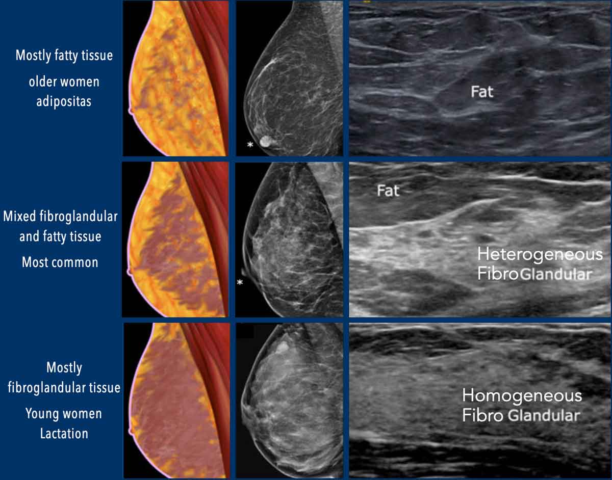
Tissue pattern
- Homogeneous echotexture – Fatty
Predominantly fatty tissue is usually seen in older women, as well as in younger or middle-aged women with obesity. - Heterogeneous echotexture
Represents a mixture of glandular and fatty tissue and is the most common breast composition pattern. - Homogeneous echotexture – Fibroglandular
A thick zone of homogeneously echogenic fibroglandular parenchyma is present beneath the isoechoic layer of subcutaneous fat lobules. Many lesions, including cancers and fibroadenomas, are found within the fibroglandular zone or at its junction with the layer of fat.
This pattern is typically seen in younger women, those with a low body mass index (BMI), and during lactation.
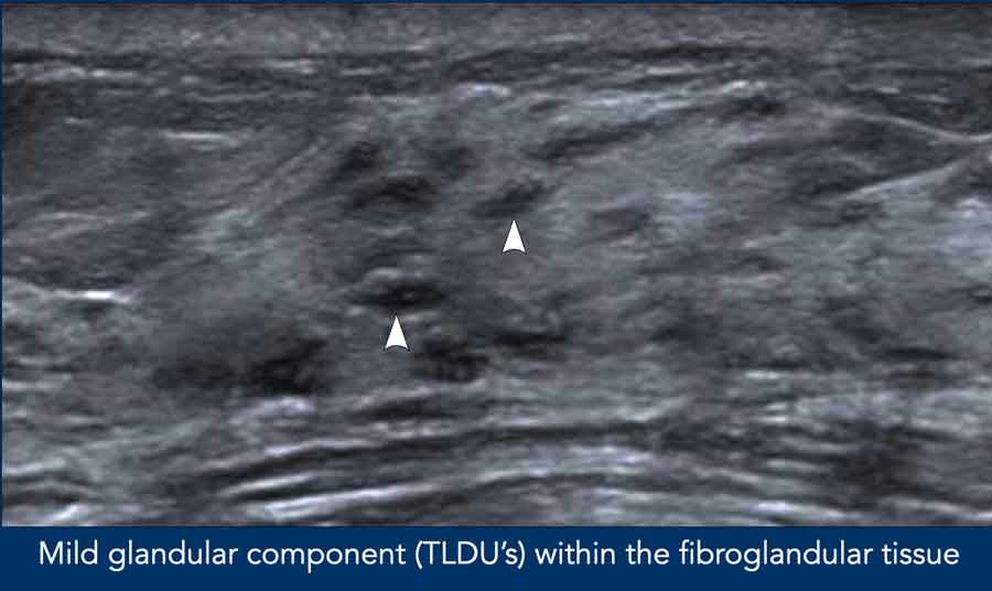
Glandular tissue component
Ultrasound can differentiate between the glandular tissue component which is the relatively hypoechoic Terminal Duct Lobular Unit (TDLU) and the stromal component, which is the hyperechoic interlobular fibrous tissue.
In one large-scale longitudinal study, an increase in the observed proportion of glandular tissue was associated with an inherent future risk of breast cancer comparable to dense breasts on mammography.
The glandular tissue component is considered:
- Minimal: < 25% of the overall fibroglandular tissue
- Mild: 25% to 49%
- Moderate: 50% to 74%
- Marked: ≥ 75% glandular tissue component.
Example
Mild glandular component.
Masses

Definition
A mass is a 3D, space-occupying lesion visible in two planes (three with volumetric imaging).

Shape
Shape
- Oval
- Lobulated - this is a new term, that is used to describe a generally oval shape with one or more indentations resulting in an undulating contour.
- Round
- Irregular

Previously the shape classification Oval (elliptical or egg-shaped) included gently lobulated masses.
In the BI-RADS v2025 Manual these are now classified as Lobulated.
Examples
- Irregular shaped IDC with echogenic rind (arrow).
- Lobulated Fidroadenoma

Margin
Circumscribed
- Sharply defined with a clear transition between the mass and surrounding tissue.
- Entire margin must be sharply defined; evaluate side margins by adjusting the transducer.
- Typically associated with oval, lobulated, or round shapes.
- Often indicates a benign mass.
- Exception: Irregularly shaped masses may occasionally have circumscribed margins, but irregular shape usually defines the mass as suspicious.
Non-circumscribed
- Indistinct - some portion of the margin is poorly defined or fuzzy
- Microlobulated - Characterized by short-cycle (2–3 mm) undulations or ripples along the margin.
Microlobulated describes the margin, while lobulated describes the shape of a mass. Microlobulations are more numerous but less deep than those defining a lobulated shape. - Angular - sharp corners forming acute angles
- Spiculated - sharp lines radiating from the mass, often a sign of malignancy
Example
Oval shaped hypoechoic mass with a microlobulated margin (arrowheads) and posterior shadowing.
Notice edema of skin and breast tissue.
Pathology: Intraductal carcinoma with mastitis carcinomatosa.
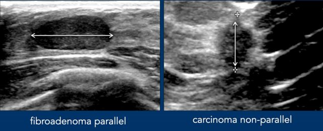
Orientation
Orientation is a feature specific to ultrasound imaging and refers to the alignment of a lesion relative to the skin surface.
- Parallel orientation (“wider-than-tall”) is typically associated with benign lesions, as the growth follows the natural tissue planes of the breast.
- Non-parallel orientation (“taller-than-wide”) is suspicious for malignancy, suggesting that the lesion is infiltrating across tissue planes.
Illustrative Cases
- Fibroadenoma: Parallel orientation (wider-than-tall).
Oval shape and well-defined margins. - Carcinoma: Non-parallel orientation (taller-than-wide).
Irregular shape and margin and echogenic rind.

Anechoic Masses
Echogenicity is an important sonographic parameter used to characterize breast lesions. It should be evaluated in conjunction with other imaging features, as echogenicity alone has limited specificity.
Anechoic echotexture typically indicates the presence of clear fluid, consistent with a simple cyst. Other anechoic structures include breast implants, postoperative fluid collections, and occasionally abscesses.
Illustrative Cases
- Multiple simple cysts in a patient presenting with a palpable lump. Only the centrally located cyst was palpable due to fluid tension; the surrounding cysts were non-palpable and isoechoic to adjacent normal tissue.
- Multiple small bilateral cysts, none of which were palpable.
- Infected cyst with hypoechoic contents and thickened wall consistent with inflammatory changes.
- Dermoid cyst connected to the skin surface.
- Anechoic cyst with intracystic mass, proven to represent intracystic carcinoma on pathology (see mixed solid and cystic masses).
- Breast prosthesis demonstrating typical anechoic appearance.

Hyperechoic masses
Hyperechoic breast masses are most often benign in nature. Common benign etiologies include hematoma, fat necrosis, fat implants, abscess, and benign neoplasms.
Silicone leakage may also present as a hyperechoic mass, frequently associated with a dirty posterior shadow.
Although rare, malignant hyperechoic lesions can occur and may correspond to invasive ductal carcinoma, invasive lobular carcinoma, lymphoma, or sarcoma.
Therefore, a hyperechoic breast lesion should not automatically be considered benign, and biopsy may be warranted in selected cases.
Many carcinomas demonstrate a hyperechoic peripheral rim, which is more extensive than the hypoechoic central component.
Illustrative Examples
- Lipoma located within the subcutaneous tissue of the breast.
- Hematoma presenting as an irregular hyperechoic region following trauma.
- Fat necrosis with a well-circumscribed post-traumatic appearance.
- Hamartoma.
- Accessory (axillary) breast tissue.
- Carcinoma showing architectural distortion with an irregular hyperechoic rim and a hypoechoic center, accompanied by posterior acoustic shadowing.
- Small carcinoma with a disproportionately large hyperechoic rim.
- Carcinoma demonstrating a hyperechoic rim.
- Silicone leakage.

Hypoechoic Masses
Hypoechoic lesions are commonly encountered on breast ultrasound and include both benign and malignant entities.
The most frequent benign hypoechoic lesions are:
- Fibroadenomas
- Infected cysts or abscesses
- Intramammary lymph nodes
- Gynecomastia
Illustrative Cases
- Fibroadenoma in a young female, showing homogeneous hypoechogenicity and smooth margins.
- Fibroadenoma with mild lobulation and posterior acoustic enhancement.
- Calcified fibroadenoma with coarse internal calcifications.
- Infected cyst with a thickened wall and internal hypoechoic debris.
- Gynecomastia: On ultrasound, may appear suspicious due to its hypoechoic fibroglandular pattern; however, mammography typically shows symmetrical retroareolar glandular tissue without suspicious features.
- Intramammary lymph node with a central echogenic hilum and a characteristic “reniform” (kidney-like) appearance, often with a visible central vessel.
- Breast abscess in a lactating woman presenting with pain, swelling, and fever. Ultrasound shows a thick-walled hypoechoic collection with surrounding skin thickening. Clinical symptoms improve after aspiration and antibiotic therapy.
- Carcinoma showing an irregular contour, angular margins, and a hyperechoic rim—features highly suggestive of malignancy.
- Carcinoma another example demonstrating similar malignant morphology.

Mixed solid and cystic masses
A mixed solid and cystic mass contains both solid (iso-, hypo-, or hyperechoic) and cystic or fluid (anechoic) components.
The important feature is the solid component, and biopsy targeting the solid component is usually required to determine the etiology of the mass. Aspiration of the cystic component may yield a false negative result.
Example
Mixed solid and cystic mass

Posterior Features
Posterior features describe the echogenicity of the tissue immediately deep to a mass and are judged relative to the echogenicity of the adjacent tissue at the same depth.
Posterior features reflect the attenuation characteristics of a mass with respect to its acoustic transmission.
Because both benign and malignant solid masses can demonstrate either attenuation (shadowing) or enhancement, posterior features are mostly of secondary rather than primary predictive value.
Note that compound imaging on modern ultrasound machines tends to reduce the conspicuity of posterior features.

Posterior Enhancement
Posterior enhancement appears as an echogenic column deep to the mass, brighter than adjacent tissue at the same depth. While it is a criterion for diagnosing simple cysts, it is not a reassuring feature for solid masses, as both benign and malignant (including high-grade carcinomas) masses may exhibit it.
Posterior Shadowing
Posterior shadowing results from ultrasound beam attenuation through the mass, creating a dark column deep to it, darker than surrounding tissue. Although often suspicious, it can occur in both benign and malignant masses. Some malignancies may present only as a shadowing band without a visible mass.
- The term "combined" has now been removed because any posterior shadowing, even shadowing behind only part of a mass, is a significant finding, that supersedes any other posterior feature.
- Refractive Edge Shadowing: A thin shadowing line behind the lateral edges of a curved mass (due to absorption/refraction) is not clinically significant and must be distinguished from deep mass shadowing.
Examples
Fibroadenoma with posterior enhancement (arrow) and hamartoma with posterior shadowing.
Occasionally, a tender breast lump may be due to focal prominent fibroglandular tissue.
On ultrasound examination, with minimal transducer pressure, this can mimic a hypoechoic mass with posterior enhancement.
However, with increased compression, the apparent lesion typically resolves, confirming that it represents normal, dense fibroglandular parenchyma rather than a true pathological mass.
Video Example:
Prominent, localized glandular tissue mimicking a hypoechoic mass with posterior acoustic enhancement.
Observe the effect of increased transducer compression.
Non-mass lesions

New in this version, the term "non-mass lesion" is introduced to describe a sonographic finding that differ from normal tissue but lack the distinct margins and shape of a mass.
Introduced by Uematsu et al. (2012), these lesions may still indicate malignancy.
The key findings are described in the table.

Example Non-mass 1
Longitudinal US image shows calcifications (arrowheads) within a segmental hypoechoic non-mass lesion.
Pathology: high grade DCIS.

The lexicon for non-mass lesions is shown in the table.
Correlation with mammography, CEM, or MRI (e.g., architectural distortion, asymmetry, suspicious calcifications, or abnormal enhancement) increases the likelihood of malignancy more than ultrasound morphology alone.
Elastography and contrast-enhanced ultrasound show potential in differentiating benign from malignant non-mass lesions.
Associated Features
Like masses, non-mass lesions may exhibit associated features.
The following features are being predictive of malignancy:
- Architectural distortion
- Posterior shadowing
- Hypervascularity on Doppler imaging
- Duct extension or abnormal duct changes
- Calcifications

Clinical Correlation
Malignancy probability rises with clinical symptoms:
- 12.5% associated with nipple discharge
- 9.1% associated with palpability
No significant correlation with patient age or mammographic density.
Histopathology
- 10–54% of non-mass lesions are malignant.
- Biopsy recommended if PPV > 2%, especially with imaging correlates or clinical symptoms.
- Common malignancies: DCIS, invasive lobular carcinoma (tubular/papillary subtypes).
- Common benign etiologies: Fibrocystic change, stromal fibrosis, fibroadenomatoid hyperplasia, sclerosing adenosis, radial scar, complex sclerosing lesion, granulomatous mastitis, intraductal papilloma, abscess, chronic mastitis, duct ectasia, atypical ductal hyperplasia, diabetic mastopathy, lymphangioma.
Example Non-mass lesion 2
Large hypoechoic non-mass lesion with a regional distribution
Pathology: Diabetic mastopathy.

Example Non-mass lesion 3
US images show a large non-mass lesion with a regional distribution.
Pathology: Granulomatous mastitis.

Example Non-mass lesion 4
US images reveal a small non-mass lesion with a focal distribution.
A biopsy was performed, and a magnetic marker was left in place.
Mammography shows no abnormalities at the site.
Pathology: DCIS grade 2

Example Non-mass lesion 5
US reveals a non-mass lesion with linear distribution on the right (white arrowheads) which proved to be a DCIS grade 2.
The mass on the left (yellow arrow) was an invasive breast cancer NST (no special type)
Calcifications

Calcifications appear as echogenic foci, particularly in hypoechoic masses or distended lactiferous ducts.
High-frequency transducers can depict both micro- and macrocalcifications, but morphology is less discernible than on mammography.
Correlation with mammography is essential to prevent misinterpretation, as not all echogenic foci are calcifications.
Clinical Significance:
- Sonography may reveal mammographically occult invasive components (mass/non-mass lesions).
- Microcalcifications on sonography may have a higher positive predictive value for malignancy than those seen only on mammography.
Ultrasound-guided biopsy followed by specimen radiography is useful for:
- Percutaneous biopsy guidance when suspicious calcifications are identified.
- Cases where stereotactic biopsy is unavailable or difficult.
Examples
Two fibroadenomas containing large calcifications.
Video Example of DCIS
Multiple microcalcifications appear as small echogenic foci within and surrounding a hypoechoic mass.
Histopathological evaluation confirmed extensive ductal carcinoma in situ (DCIS), with a central focus of invasive carcinoma.
Associates Features

Effects of a pathologic process on its surroundings may include
- Architectural distortion like tissue compression, Cooper’s ligament thickening, ductal aberrations, or echogenic pseudocapsule or echogenic rind.
- Additional findings like breast edema, skin thickening (due to inflammatory cancer, radiation, mastitis, or systemic conditions like congestive heart failure).
- Doppler/Elastography: Abnormal vascular findings or tissue stiffness.
These findings can help to characterize a lesion and distinguish malignant from benign lesions, but the morphology of a lesion supersedes associated features in management decisions; suspicious morphology still requires biopsy.
Echogenic Pseudocapsule
- Definition: Thin, uniform white line surrounding a mass’s margin.
- Benign Likelihood: Oval or lobulated masses with a circumferential pseudocapsule and no suspicious features are very likely benign (consider interval growth).
- Biopsy Indication: Round or irregular masses should be biopsied regardless of pseudocapsule presence.
Echogenic rind
Definition
A thick echogenic band surrounding all or part of a breast mass or non-mass lesion, disrupting normal tissue texture. Likely represents a desmoplastic reaction or peritumoral edema.
Clinical Significance
- High predictive value for malignancy (though it can accompany benign findings).
- Suspicious feature unless imaging confirms a definitively benign cause (e.g., fat necrosis, infection, hematoma).
- Measurement: Include the rind when measuring mass/non-mass lesion size for accurate histologic correlation.
Video example
Thick irregular echogenic rind surrounding an irregular hypoechoic mass. Pathology: intraductal carcinoma.

Edema
Edema is haracterized by interconnected, arborizing hypoechoic or anechoic lines, representing dilated lymphatics or interstitial fluid.
Often accompanied by skin thickening.
Unilateral Edema
- Suggests a focal process (e.g., inflammatory breast cancer, mastitis, post-radiation changes).
Bilateral Edema:
- Suggests a systemic disorder (e.g., congestive heart failure, renal failure, central venous obstruction).

Skin thickening / retraction
Definition
- Skin thickness > 2 mm (up to 4 mm in the peri-areolar area is normal).
- Subtle thickening is confirmed by asymmetry compared to the contralateral breast.
- Often involves loss of the two echogenic lines that define the normal skin layer.
Example
- Ill-defined hypoechoic retromammillary mass in a breastfeeding woman.
- Symptoms: Painful swelling with thickened, red skin (indicated by arrow).
- Diagnosis: Abscess. Percutaneous drainage through thick needle aspiration.

Vascularity
Evaluated using power and color Doppler.
Vascular patterns support diagnoses but are not sole diagnostic criteria.
Note: No vascular pattern is specific for any diagnosis; both benign and malignant masses may show increased, minimal, or no vascularity.
Avascular
- Common Example: Cysts.
- In solid masses, absence of vascularity suggests little or no blood flow.
- Transducer pressure should be minimal to avoid occluding small vessels and falsely suggesting avascularity.
Internal vascularity
- Typically confirms a solid mass or solid components.
- In Non-Mass Lesions:
- Focus on the degree of vascularity compared to normal breast tissue or the contralateral breast.
- Expectation: Non-mass lesions generally show increased vascularity relative to surrounding normal tissue.
Peripheral vascularity
- Tissue surrounding the abnormality is hyperemic, possible due to inflammation, infection,or neovascularization
Special Cases

Special cases are those with a unique diagnosis or pathognomonic findings.
Breastimplants are discussed in the article "Breast implants - normal findings and complications".

Clustered microcysts
Description
Aggregation of anechoic masses with thin (<0.5 mm) septations and no discrete solid component.
Observed in up to 6% of ultrasound examinations.
Classic criteria: individual cystic spaces <2–3 mm; however, malignancy risk does not increase if size criterion is extended to 7 mm. Margins may appear microlobulated due to small cysts but should remain distinct.
Associated Diagnoses
Fibrocystic changes, apocrine metaplasia.
Typically benign (BI-RADS category 2), especially if multiple or associated with fibrocystic changes. BI-RADS 4 (suspicious) if margin is indistinct or a discrete solid component is present; biopsy recommended, particularly for new masses or in post-menopausal patients.

Complicated cyst
Description
Cysts containing debris, without a discrete solid component, and with a thin wall.
Debris may appear as diffuse, homogeneous low-level echoes, sometimes layered, shifting slowly with patient position changes, or as diffuse mobile scintillating reflectors.
Example
A painful hypoechoic lesion was identified. Doppler ultrasound demonstrated no internal vascularity. The differential diagnosis included a complicated cyst versus a solid lesion. Percutaneous aspiration yielded purulent material, confirming a complicated cyst.

Example
A complicated cyst filled with debris.
By turning the patient to the side a fluid-fluid level became visible.
There was no intracystic tumor.

Mass in or on skin
Description
Clinically apparent masses, including sebaceous or epidermal inclusion cysts, keloids, moles, pimples, neurofibromas, and accessory nipples.
Skin metastasis are rare, but can be seen in the context of a mastectomy scar; clinical history of a primary tumor aids interpretation.
Confirm the mass is at least partially within the two thin echogenic bands of the skin.
Examples
Two intradermal lesions surrounded by some inflamed subcutaneous fat.

Fat necrosis
Fat necrosis is related to surgical or non-surgical breast trauma.
In the acute phase there is local edema on ultrasound (images), progressing to a mixed solid/cystic mass, complicated cyst, or anechoic cyst within 2–6 months.
In the chronic phase (1–2 years): May evolve into a calcified oil cyst or spiculated mass, both with posterior shadowing; the latter is more often associated with surgical trauma.
Diagnostic Considerations
- US appearance is variable and may mimic suspicious findings.
- Correlation with mammography is essential, as oil cysts (solitary/multiple, calcified/non-calcified) are typically benign on mammography but not necessarily on US.
- Recommendation: If fat necrosis is suspected on US, correlate with mammography to confirm a benign (BI-RADS category 2) assessment, as US alone may be insufficient.
Lymph nodes

Axillary nodes
Normal lymph nodes are typically oval or reniform with a circumscribed margin. They consist of a hypoechoic cortex and an echogenic hilum containing fat. The presence of flow in the hilum aids in their identification.
Parameters for Lymph Node Characterization
- Size
- Shape
- Oval or reniform
- Lobulated
- Round
- Irregular
- Cortical Thickening (> 3 mm)
- Diffuse, concentric, uniform.
- Focal, eccentric: Hypoechoic cortical bulge may indicate nodal metastases, with tumor cells trapped in cortical follicles.
- Margin
- Circumscribed
- Non-circumscribed: Indistinct margins may suggest extranodal tumor extension.
- Hilum
- Pathological nodes may show diminished or eccentrically located hilar fat.
- Complete hilar effacement suggests a high probability of malignancy.
Examples
A normal axillary lymph node and a focal metastasis (proven by US-guided cytologic puncture)

Clinical Correlation of lymph nodes
Breast cancer primarily metastasizes via lymphatic spread, so abnormal nodes on ultrasound should raise suspicion for malignancy.
However, benign etiologies (e.g., autoimmune diseases, connective tissue disorders, granulomatous disease, lymphoma, or vaccine-related adenopathy—such as post-COVID-19, influenza, pneumococcus, or shingles vaccinations) must also be considered.
Example
A patient presented with unilateral enlarged axillary lymph nodes.
Ultrasound examination of the breast reveiled a small intraductal carcinoma, not visible on mammography, because it was hidden in dense fibroglandular tissue.

Intramammary nodes
Intramammary lymph nodes are commonly located in the upper outer quadrant and posterior third of the breast (particularly the axillary tail), often adjacent to blood vessels.
Their typical size ranges from 3–4 mm to 1 cm, though they may be larger.
Due to their small size, the echogenic fatty hilum may be difficult to visualize.
Doppler imaging can help identify the hilum by detecting the afferent artery and efferent vein.
Breast cancer can metastasize to intramammary lymph nodes, which may then appear enlarged, round, or irregular.
Metastases to intramammary lymph nodes are staged equivalently to axillary level I nodes in the TNM classification.
Example
Small oval shaped well-defined hypoechoic mass in the right breast suspicious for a normal intramammary lymph node. No fatty hilum is visible.
Color doppler reveales the afferent artery and efferent vein.
Location of findings

The location of a breast finding should be described to ensure easy identification and correlation with clinical and imaging findings.
- Laterality: Always specify left or right breast.
- Position in Breast: Use clock face (preferred) or quadrant for location.
- Clock face: Oriented with the patient facing the observer; use whole numbers (e.g., 3:00 = lateral left breast, medial right breast).
- Quadrant: Use only for large findings spanning multiple clock face locations (e.g., upper outer, upper inner, lower outer, lower inner).
- Distance from Nipple: Report as whole centimeters from the nipple base to the finding’s center (e.g., 2 cm from the nipple). For large findings, include distances to anterior and posterior extents.
- Anatomic Zone: Optionally, describe the zone (e.g., subcutaneous fat-fibroglandular junction) to aid multimodal correlation.
Example Description: Right breast, 3:00, 2 cm from the nipple, at the subcutaneous fat-fibroglandular junction.
Measurements of findings

Rounding:
- Round measurements to one decimal place (cm) or whole numbers (mm).
- Example: 0.45–0.49 cm → 0.5 cm, 2.43 cm → 2.4 cm, 12.4 mm → 12 mm.
- Maintain consistent units throughout the report.
Lesion Measurements:
When possible, provide three measurements:
- Longest axis,
- Perpendicular to the first,
- Orthogonal to the first two (representing the unmeasured plane).

In mammography, the short axis of a lymph node is measured.
In ultrasound, assessment focuses on focal cortical thickening with potential tumor deposition.
Assessment Categories
