32 cases of suspected COVID-19
Imaging findings and follow up
Frank Smithuis and Robin Smithuis
Academical Medical Center Amsterdam and Alrijne Hospital Leiderdorp, the Netherlands
Publicationdate
The role of CT in this COVID-19 pandemic still has to be determined.
CT can help to determine the severity of the disease and is a valuable and fast tool to determine whether a patient is infected by COVID-19 or not.
This can help to keep patients with a high suspicion of COVID-19 infection separate from patients with other diseases, especially when the PCR-test is false negative.
These cases are all patients that were admitted to the hospital with suspicion of COVID-19 infection and all were PCR-tested.
The findings at presentation and follow up is provided.
In the description we use the CO-RADS classification.
Press ctrl+ for larger images and text on a PC or ⌘+ on a Mac.
This can be helpful for scroll-images.
Single images can be enlarged by clicking on them.
Introduction
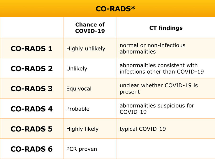
The CO-RADS classification is a standardized reporting system for patients with suspected COVID-19 infection developed for a moderate to high prevalence setting.
It is a preliminary classification of likelyhood of COVID-19 infection as proposed by the COVID working group of the Dutch Radiological Society.
The interpretation of the CT findings has to be combined with the clinical symptoms and the duration of the symptoms as a CT can be negative in the first few days of a mild infection.
However most patients that we see have complaints for a week or more.
At the moment most patients that are admitted to the hospital either have a CORADS 5, which means that they have a COVID-19 infection or they have a CORADS 1 or 2, which means no COVID-19 infection.
Imaging Findings in suspected COVID-19
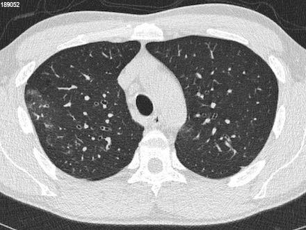
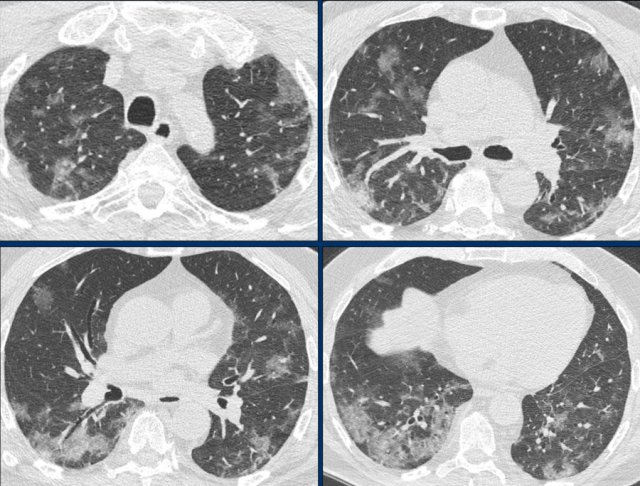
_1 Crazy paving - ventilation
History
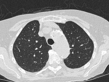
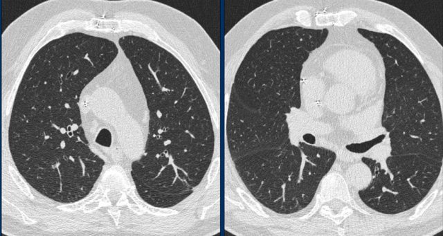
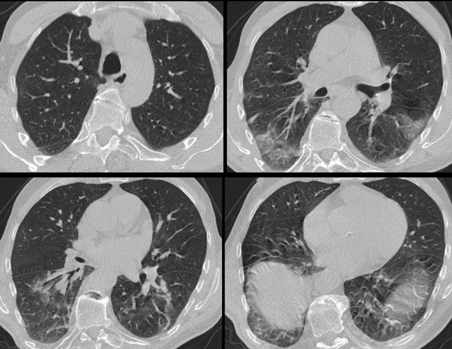
64 year old male with fever and coughing for 2 weeks after a skiing holiday with his family.
CT findings
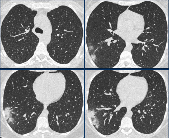
Widespread GGO in all lobes
Crazy paving (blue arrows)
Vascular enlargement (black arrow)
Subpleural bands with retraction (yellow arrows)
Consolidation and bronchiectasis posteriorly in the lower lobes
CORADS 5 - very high suspicion of COVID-19
PCR positive
Continue with Chest X-ray...
Within a few hours after presentation on the ER the patient became hypoxic and was treated with mechanical ventilation.
Later that day the patient was transferred to another hospital.
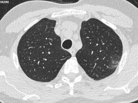
_2 CORADS 5 - extubation
History
55 year old and previously healthy man presented with a history of 2 weeks of fever and coughing. Although he was in a reasonable condition at arrival, he had to be intubated later that evening.
CT findings at arrival
Consolidations mainly posteriorly in lower and upper lobes
Small areas of GGO
CORADS 5
PCR
The first PCR was negative, but later a sputum test was positive for COVID-19.
Follow up
After three days of mechanical ventilation he could be extubated and was doing well with only oxygen therapy. Nine days after admission to the hospital he was discharged and is doing well.
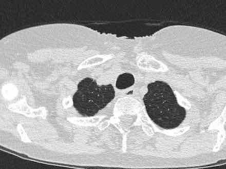
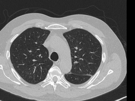
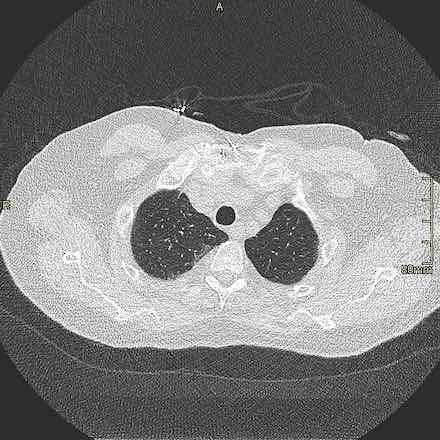
_3 CORADS 2 - Mycoplasma
History
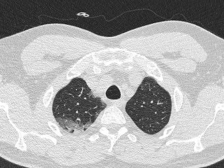
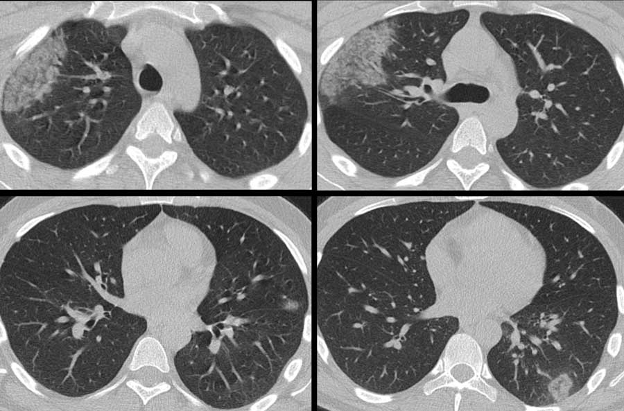
49 year old male suspected of having COVID-19.
13 days of fever and coughing. Treated with antibiotics for 7 days.
CT findings
Consolidation in right lower lobe
surrounding area with tree-in-bud in lower lobe
also tree-in-bud in other lobes
CORADS 2 - some other infection most likely bacterial
PCR
First test negative. Test nine days later also negative.
Follow up
Tested positive for Mycoplasma pneumoniae. Left the hospital two days after admission.
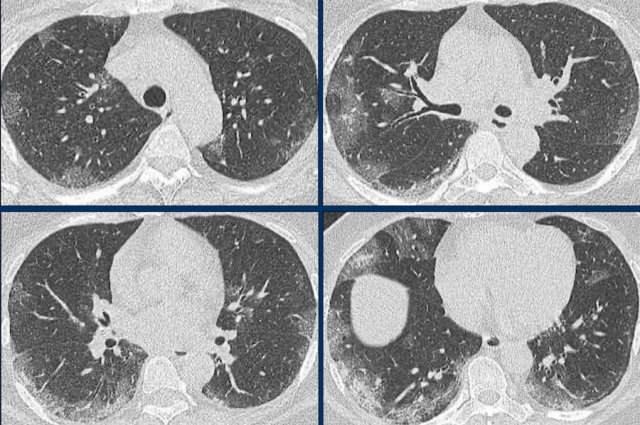
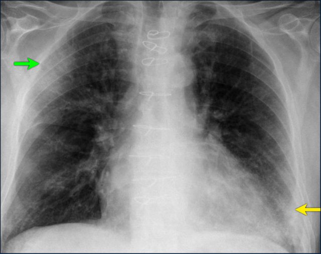
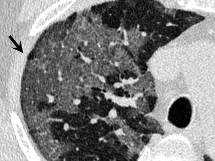
_4 GGO - bronchiectasis- wide vessels
History
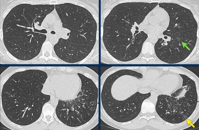
75 year old male with fever for 4 weeks and no coughing. History of lungcancer resection by video-assisted thoracoscopic surgery (VATS) one years ago.
CT findings
Bilateral GGO
Bronchiectasis (green arrow)
Widened vessels (yellow arrow)
CORADS 5
PCR
2x positive
Follow up
Two days after admission to the hospital, there was a rapid decrease in condition of the patient and he had to be transferred to the ICU for mechanical ventilation.
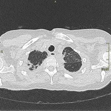
_5 CORADS 2 - Asthma
64 year old female known with asthma suspected of having COVID-19.
CT findings:
- Mucus plugging in right lower lobe bronchus
- Consolidation and atelectasis of right lower lobe
- Consolidation in middle lobe
CORADS 2 - infection not related to COVID-19
PCR: negative for COVID-19 (twice) and negative for RSV, Inflluenza A and B.
 Click image to enlarge
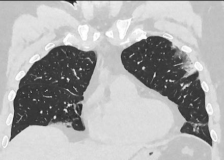
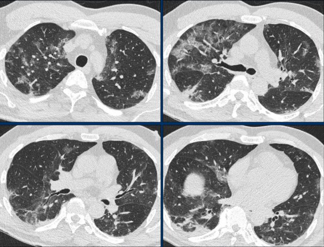
_6 CORADS 5 - bilateral peripheral GGO
COVID-19 infection.
CT-findings:
- Predominantly bilateral subpleural GGO with some areas of crazy paving.
- In the lower lobes some areas of consolidation.
- Percentage of lung involvement is approximately 25% by visual assessment.
- CORADS 5
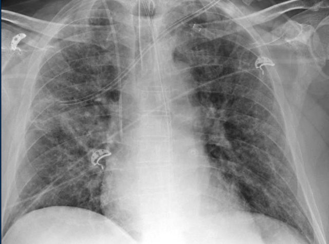
_7 Fatal COVID-19
83 year old male with mitral insufficiency and pulmonary hypertension was diagnosed with COVID-19 infection.
The chest film shows consolidation in the right upper lobe (green arrow) and probably some consolidation in the left lower lobe.
The patient decided not to be treat with mechanical ventilation and died four days later.
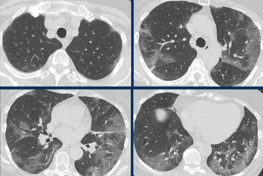
_8 CORADS 5 - Crazy paving
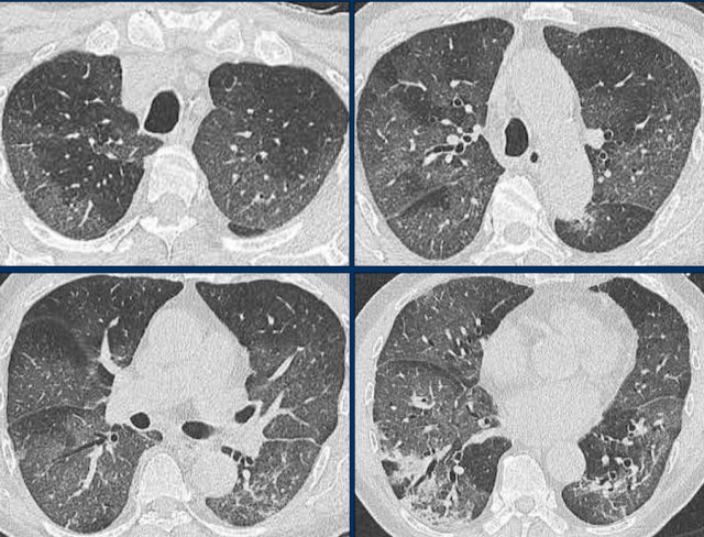
57 year old male without any prior diseases was admitted to the hospital with 14 days of fever and cough.
He was treated with an oxygen mask.
2 days later his condition suddenly worsened and the patient was tranferred to the ICU for mechanical ventilation.
CT findings at the moment of admission to the hospital:
- Bilateral GGO
- Crazy paving with thickened intra- and interlobular septa
- CORADS 5 - typical COVID-19
PCR: positive for COVID-19.
Crazy paving, consolidation, linear opacities, bronchial wall thickening and high CT scores are features of severe and critical COVID-19 pneumonia (1).
_9 Suspicion pulmonary emboli
57 year old male with Diabetes type 1 with chronic obstructive lung disease was admitted with shorteness of breath.
Initially there was no suspicion of COVID-19.
A CT was performed to look for pulmonary emboli.
CT findings:
- No emboli
- Bilateral GGO with a centrilobular pattern imposed
- CORADS 3 - unsure COVID-19
PCR was two times negative for COVID-19 and all other test for RSV, influenza, legionella and pneumococcus were also negative.
The patient was treated with an oxygen mask for 7 days and then recovered.
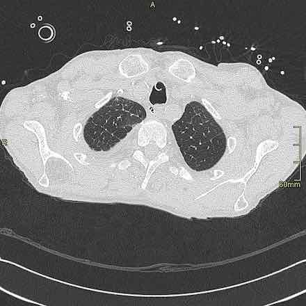
10 CORADS 5 - Subpleural bands
69 year old female with mild dyspnoe for one week with cough and fever.
She was treated with 2L O2/min.
CT findings:
- Subpleural bands and some GGO
- CORADS 5
11 CORADS 1 - Colitis
History
89 year old female, who had fever for seven days with diarrhoea.
CT findings
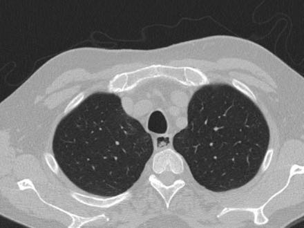
Normal lungs
Thickened wall of the descending colon probably colitis
CORADS 1 no COVID-19
PCR ...
12 CORADS 5 - Peripheral consolidation
49 year old male complained of being extremely tired for 12 days with headache and a dry cough and weight loss of 8 kilos.
CT findings:
- Bilateral consolidation more pronounced in the lower lobes and peripheral.
- The consolidations are more pronounced then the GGO
- CORADS 5
He was treated with an oxygen mask for 3 days and then recovered.
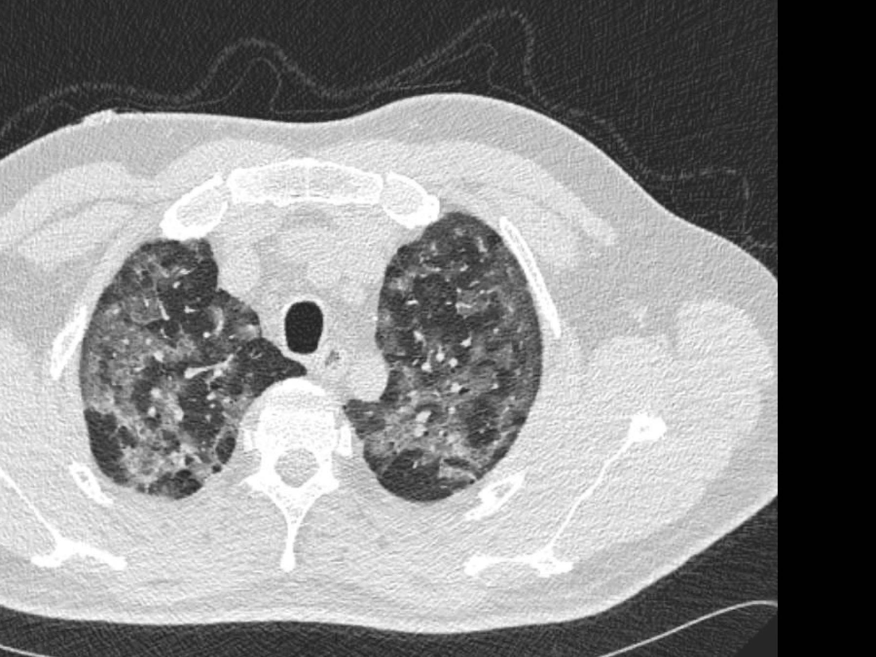
13 CORADS 5 - 75% involvement
History
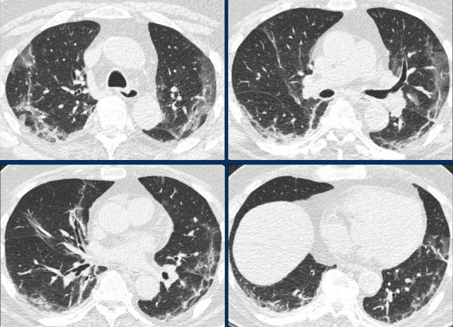
40 year old male, who had fever for ten days with progressive coughing and shortness of breath. Saturation at admission was 66%.
CT findings
Widespread bilateral ground-glass opacities with a posterior predominance.
75% of the lungs are involved.
CORADS 5 very likely COVID-19
PCR positive
14 Subpleural bands
History
75 year old male with fever for 6 days
CT findings
Bilateral subpleural bands
CORADS 5
Comment
Subpleural bands are probably fibrous bands but this is still not certain.
Pan reported 17% COVID-19 patients with fibrous stripes in their study (2).
Fibrous lesions may form during the healing of pulmonary chronic inflammation or proliferative diseases, with gradual replacement of cellular components by scar tissue.
The relation between fibrosis and patients’ prognosis is debatable.
 Click to enlarge
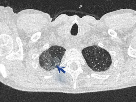
15 CORADS 3 - focal consolidation with halo
History
34 year old female
High fever for 1 day with coughing
CT findings
Focal consolidation with surrounding GGO
Only in right lower lobe.
CORADS 3 equivocal
PCR 2 x negative
Influenza negative, RSV negative
Clinical course
Continuous fever for two more days. No oxygen. Discharged from hospital on third day.
16 Mild infection
History
61 year old male
10 days fever, dyspnoe and diarrhoea after a holiday in Egypt.
CT findings
Bilateral faint areas of GGO
Severity index: 5
10% involvement
CORADS 5 very likely COVID-19
PCR positive
Clinical course
After 2 days of oxygen therapy the patient could be discharged from the hospital
17 GGO only in right lung
History
46 year old male
8 days fever, dry cough, dyspnoe and diarrhoea.
CT findings
GGO in the right lung
Severity index: 5
10% involvement
CORADS 5 very likely COVID-19
PCR not known
Clinical course
not known
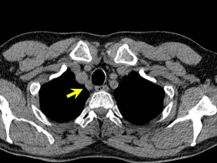
18 Mild lymphadenopathy
History
61 year old male had high fever for 10 days.
CT findings
Bilateral patchy areas of GGO
CORADS 5 very likely COVID-19
PCR positive
Clinical course
4 days of Oxygen therapy
19 Lobar consolidation and GGO
History
67 year old male, who had fever for fourteen days with coughing and lately hemoptoe.
CT findings
Dense consolidation in left lower lobe
Ground glass in right lower lobe (yellow arrows)
Maybe some tree-in-bud in right upper lobe (red arrow)
CORADS 2 low suspicion COVID-19, probably bacterial pneumoniae
PCR 2 x negative
Influenza negative, RSV negative, no pneumococcus, no legionella.
Treated with antibiotics and was feeling better 2 days later with no fever.

20 Extubation
History
73 year old male with aorta insufficiency and pacemaker was admitted to the hospital with fever and coughing after being in an area with Corona.
X findings
day 1. normal findings
day 4. bilateral consolidations intubated.
day 8. bilateral consolidation
day 13. extubation
PCR
positive
Follow up
Extubated after 9 days of mechanical ventilation
21 Bilateral patchy GGO - no oxygen
History
71 year old male coughing for 10 days, no fever
CT findings
Bilateral patchy areas of GGO
CORADS 5 very likely COVID-19
PCR positive
Clinical course
Did not need oxygen therapy. Discharge 4 days later.
22 Bilateral GGO 3 days oxygen
History
61 year old male with fever, coughing for 1 week.
CT findings
Bilateral patchy GGO
CORADS 5
PCR
positive
Follow up
Discharge after 3 days of oxygen therapy

23 Vacuolar sign
History
67 year old woman was admitted to the hospital after spending one week in quarantaine with fever, coughing and headache.
CT findings
Patchy areas of GGO bilateral
Bronchiectasis
Wide vessels
Vacuolar sign (1)
Subpleural bands in lower lobes
CORADS 5 very likely COVID-19
PCR positive
Clinical course
One day after admission she was intubated and transported to another hospital.
24 Fever and hemoptoe
History
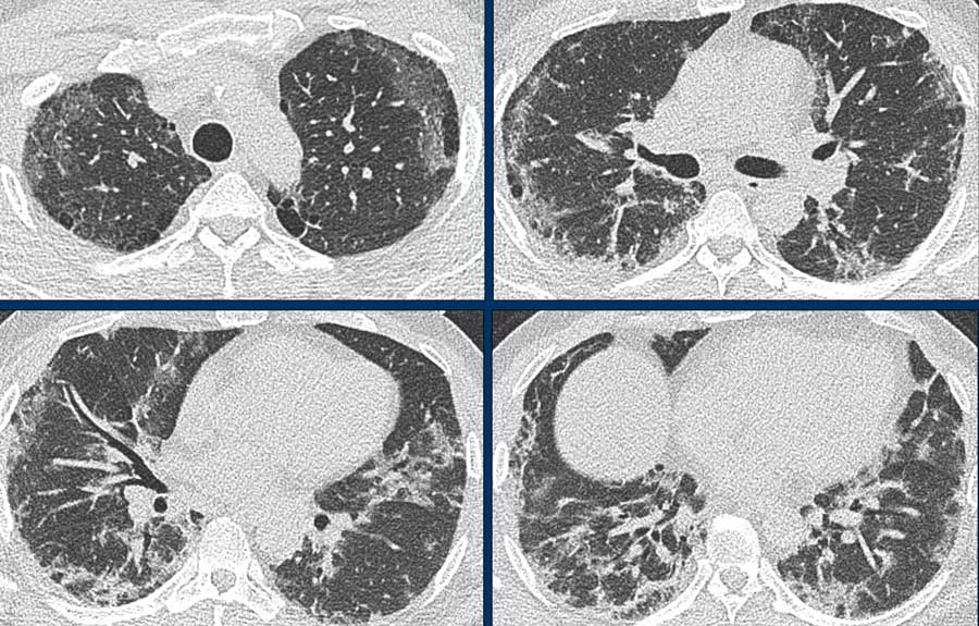
58 year old male with mild mitral and aortic insufficiency presented with high fever and coughing for 10 days with exhaustion.
CT findings
Extensive bilateral GGO
Extensive widenend vessels
Bronchiectasis
> 75% lung involvement
CORADS 5
PCR
positive
Follow up
Immediately after admission the patient was transferred to the ICU and intubated. Patient died eight days later.
25 CORADS 1 Coughing and chest pain
History
61 year old male with a history of bypass surgery and endocarditis complicated by a total AV block for which he had a pacemaker, presented with coughing for 1 week and chest pain.
CT findings
Normal
CORADS 1
PCR
negative - results after discharge
Follow up
There was no cardiac problem involved. Because of the normal CT the patient was reassured and returned to his home and he received a later call that the PCR was negative.
 Click image to enlarge
26 Immunodeficiency
History
50 year old female with a common variable immunodeficiency (CVID) had complaints of a cold with a non productive cough was admitted to the hospital because she had fever for one day and headache.
CT findings
Subtle findings only in left lower lobe
Septal thickening
Subtle areas of GGO
Bronchial wall thickening
CORADS 3 indeterminate
PCR negative
 Click image to enlarge
27 CORADS 5
History
47 year old male with flew-like symptoms for 10 days was admitted to the hospital with progressive dyspnoe and an oxygen saturation of 82%.
CT findings
Bilateral peripheral GGO
Bronchiectasis
CORADS 5

28 CORADS 4
History
40 year old female presented with acute dyspnoe and hemoptoe
CT findings
Areas of GGO and basal consolidation in lower lobes.
CORADS 4
probable COVID-19
PCR
Positive
29 RSV infection
History
67 year old male with Non Hodgkin Lymphoma who had a allogeneic stem cell transplantation half a year ago, was admitted to the hospital with high fever and cold shivers since one day. No coughing.
CT findings
Multifocal consilidations with halo sign
CORADS 3
equivocal COVID-19
PCR
Negative. RSV positive
30 CORADS 5 subpleural bands
History
79 year old male presented with one week dyspnoe and non productive coughing. Received antibiotics since two days. Since one day high fever. R
CT findings
Bilateral GGO
subpleural bands
CORADS 5
very likely COVID-19
PCR
positive
31 Chest pain and low body temperature
History
63 year old female presented with dyspnoe and chest pain since one day. She had a low body temperature and low oxygen saturation. ECG and troponines were normal.
CT findings
No pulmonary emboli or dissection
Some GGO and consolidation not the bilateral patchy pattern that we normally see in COVID-19.
Thickened interlobular septa
Pleural fluid
CORADS 2
low suspicion of COVID-19
Maybe some other infection in combination with heart failure.
PCR
negative
32 probably heart failure
History
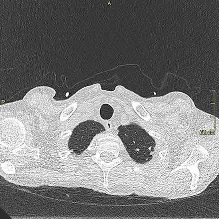
56 year old male with a history of two times renal tranplant with rejection and hypertension and vomiting and diarrhoea since three days. At admission the oxygen saturation was 74%.
CT findings
high position tube
diffuse GGO and thickened interlobular septa
bilateral pleural fluid
CORADS 2
Low suspicion of COVID-19. Most likely heart failure with pulmonar edema.
PCR
Negative. Influenza and RSV negative
33 CORADS 5
History
73 year old female known with LBTB had progressive dyspnoe for 3 weeks.
CT findings
Bilateral widespread areas of GGO
CORADS 5
typical COVID-19
PCR
positive
34 CORADS 3 PCR+
History
70 year old male with dementia was admitted to the hospital with chest pain and dyspnoe. No cardiac cause.
CT findings
breathing artifacts.
maybe some areas with GGO
CORADS 3
Indeterminate
PCR positive
35 25 year old male
History
25 year old male with fever and dyspnoe for 5 days. Treated with oxygen for one day. Went home but was readmitted the next day with progressive dyspnoe.
CT findings
Bilateral GGO
CORADS 5
PCR positive