Normal Values in Pediatric Ultrasound
Simon Robben, Rick van Rijn and Robin Smithuis
Radiology Departement of the Maastricht University Hospital, Academical Medical Centre in Amsterdam and the Alrijne hospital in Leiderdorp, the Netherlands
Publicationdate
This is an overview of normal values of ultrasound examinations in neonates and children.
Click on one of the items on the left.
Adrenal
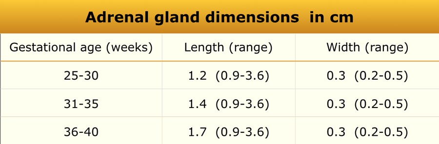 Adapted from reference 21
Material and methods
Ultrasonographic study of 92 infants.
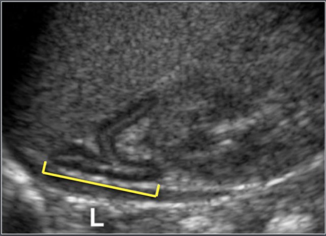
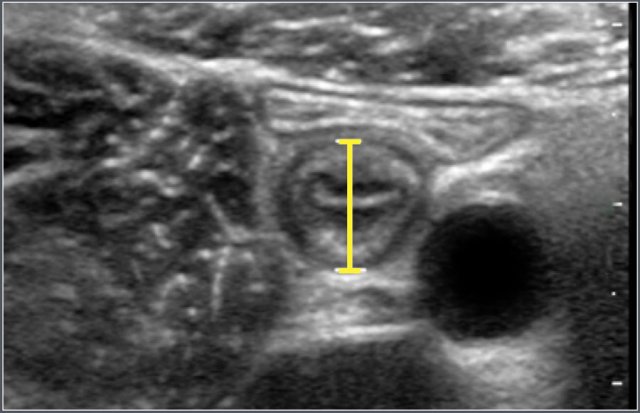
Sagittal measurement of the adrenal gland.
Causes of enlargement of adrenal glands:
- Congenital Adrenal Hyperplasia
- Adrenal Hemorrhage
- Adrenal Neuroblastoma
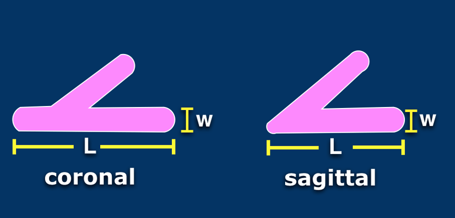
The length (L) of the gland is defined as the maximum cephalocaudal dimension (either coronal or sagital plane).
The width is defined as the maximum thickness of one of the limbs.
Appendix
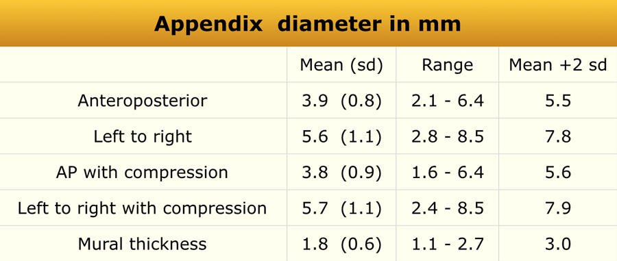 Adapted from reference 14
Materials and method
In this ultrasonographic study 146 consecutive patients (62 boys and 84 girls; mean age, 7 years; age range, 2-15 years) were included.
Children with cystic fibrosis, acute abdominal pain, with previous appendectomy and below the age of 2 years (because of difficulty in performing the examination) were excluded.
In 120 children the appendix was visualized.
Ultrasonographic anteroposterior measurement of the appendix.
Casues of enlarged appendix:
- Appendicitis
- Cystic fibrosis
- Lymphoid hypertrophy (Immune deficiency, viral enteritis)
- Intraluminal gas, mucus of faeces
- Mucocele
Bladder
 Adapted from reference 17
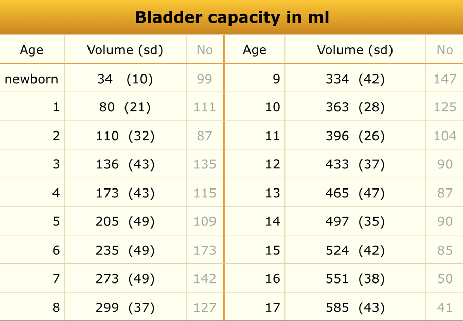
Bladder volume
Material and method
A total of 3376 children were recruited in this ultrasonographic study.
The total number of patients does not add up to the total number of patients in this study because not all age-subgroups were included in the table.
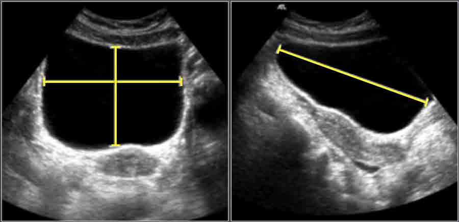
The bladder volume was calculated first by measuring the maximum length (L) of the urinary bladder on the longitudinal scan, which was obtained from the neck to the fundus of the bladder.
Depth (D) was measured, perpendicular to the first plane at the level of the maximum area, in the midline from the anterior to posterior mucosal surface of the bladder.
The width (W) was taken perpendicular to D at its mid-point.
Bladder volume as presented in the table was recalculated from the data in this study using the equation for an ellipsoïd: L×D×W (in centimetres) x 0.523.
 Adapted from reference 17
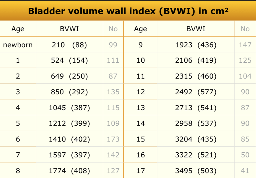
Bladder wall thickness
Materials and method:
A total of 3376 children were recruited.
Bladder wall thickness was only measured when the residual bladder volume was <10% of the original volume.
Causes of bladder wall thickening:
- cystitis
- dysfunctional voiding
- urethral valves
The bladder wall thickness was measured from a zoomed image of the transverse plane of the voided bladder at 3 points: anterolaterally, laterally and posterolaterally (figure). The mean was taken for these three measurements.

The bladder wall thickness depends on the degree of filling of the bladder and its capacity.
Therefore the bladder wall thickness is expressed as the bladder volume wall thickness index (BVWI).
Bowel
 Adapted from reference 13
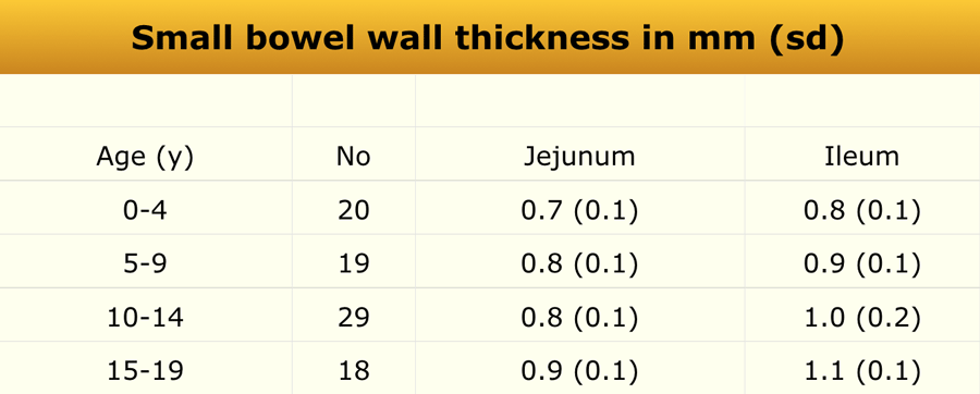
Materials and method
The study population consisted of 128 patients (57 male and 71 female).
Of this population 86 were between the ages of 1-19 years (only data pertaining to this selection is presented).
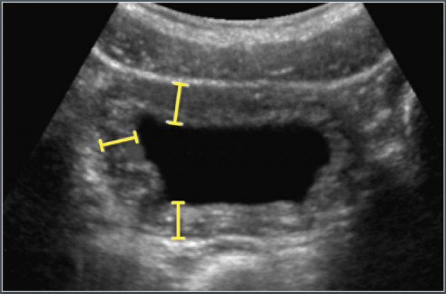
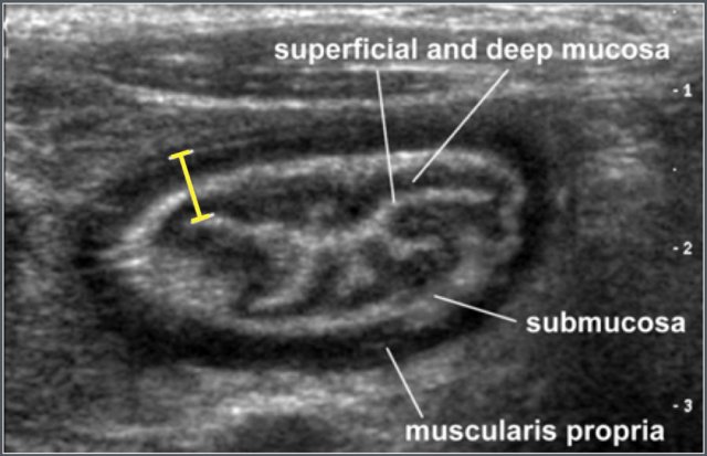
Bowel wall thickness was measured on transverse sections and comprised of mucosa, lamina propria, muscularis mucosa, submucosa, and muscularis propria.
Ultrasonographic measurement of wall thickness of terminal ileum in a 12-year-old boy with cystic fibrosis.
Causes of small bowel wall thickening:
- Henoch Schönlein Purpura
- Crohn's disease
- Lymphoma
 Adapted from reference 13
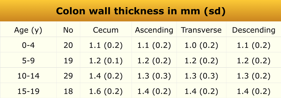
In the same study the wall thickness of the colon was measured.
Causes of colon wall thickening:
- Inflammatory bowel disease (IBD)
- Hemolytic Uremic syndrome
- Pseudomembranous colitis
Common Bile Duct
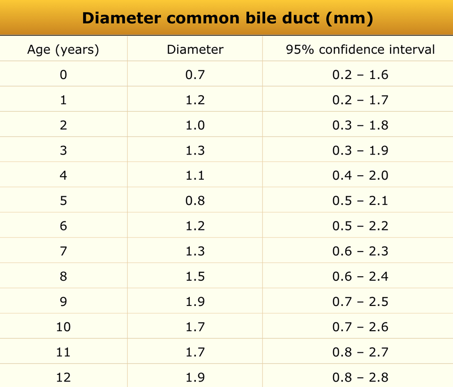 Adapted from reference 8
Materials and method
One hundred and seventy-three consecutive children, referred for abdominal ultrasonography not related to hepato-biliary pathology, were included in this study (100 boys and 73 girls), age range 1 day - 13 years (median age 5.0 years).
The diameter of the common bile duct was ≤ 3.3 mm in all patients.
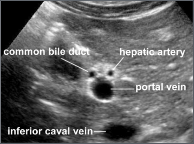
Transverse ultrasonographic image of common bile duct and surrounding anatomy
Galbladder
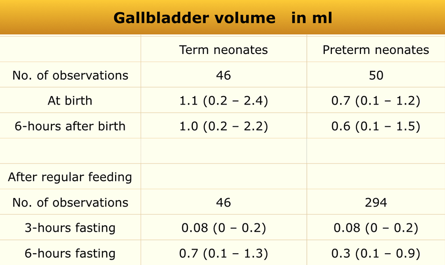 Adapted from reference 10
Materials and method
Ultrasonographic gallbladder volume assessment (length x width x height x 0,52) was performed in 50 preterm (mean GA 31.7 ± 2.5 weeks and mean birth weight 1556 ± 441 g) and 46 term infants (mean GA 38.3 ± 1.2 weeks 3253 ± 440 g).
Data were collected soon after delivery and at 6-h fasting, and at the age of 5-7 days at 3-h and 6-h fastening following regular milk feeding.
Causes of small gallbladder:
- Biliary atresia
- Fatty meal
Hip
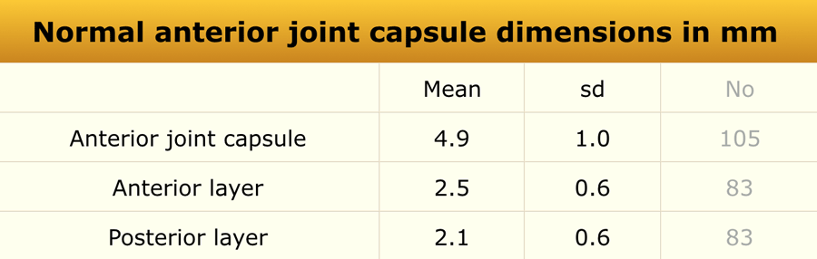 Adapted from reference 22
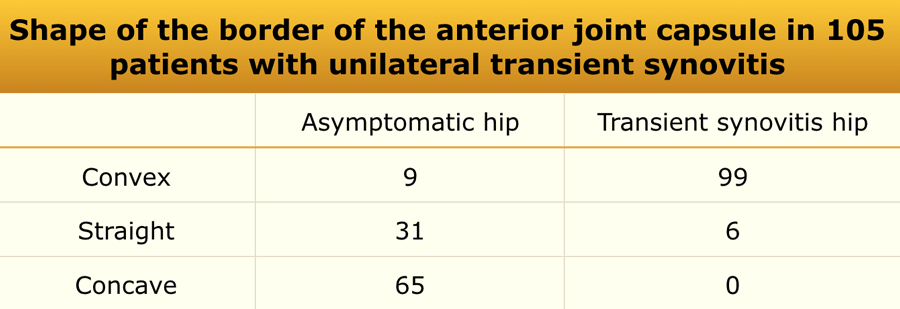
Anterior recess
Materials and method
Ultrasonographic study of 58 healthy children and 105 children with unilateral transient synovitis (age range 1.7-12.8 years).
The children were examined in the supine position with hips in neutral position.
 Adapted from reference 22
The children are examined in the supine position with hips in neutral position.
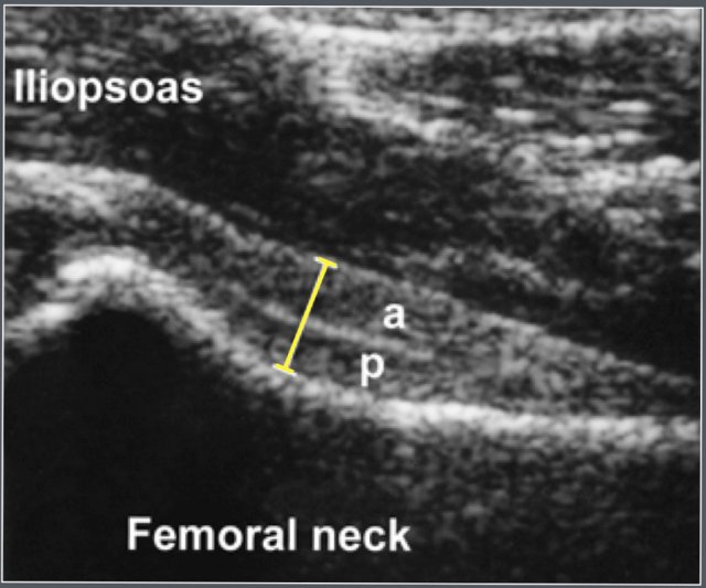
The anterior joint capsule was measured, including both of its components (the anterior and posterior layer).
Also the anterior contour of the joint casule is evaluated.
There was no statistically significant correlation between age and thickness of the anterior joint capsule.
A difference >2mm or an effusion >2mm is considered abnormal.
Shape of the border of the anterior joint capsule
The anterior contour of the joint capsule can be evaluated.
Ultrasonographic measurement of the anterior joint capsule. Both anterior (a) and posterior (p) layer can be identified.
Causes of hip joint effusion:
- Transient synovitis
- Septic arthritis
- Juvenile Idiopathic Arthritis
 Adapted from reference 23
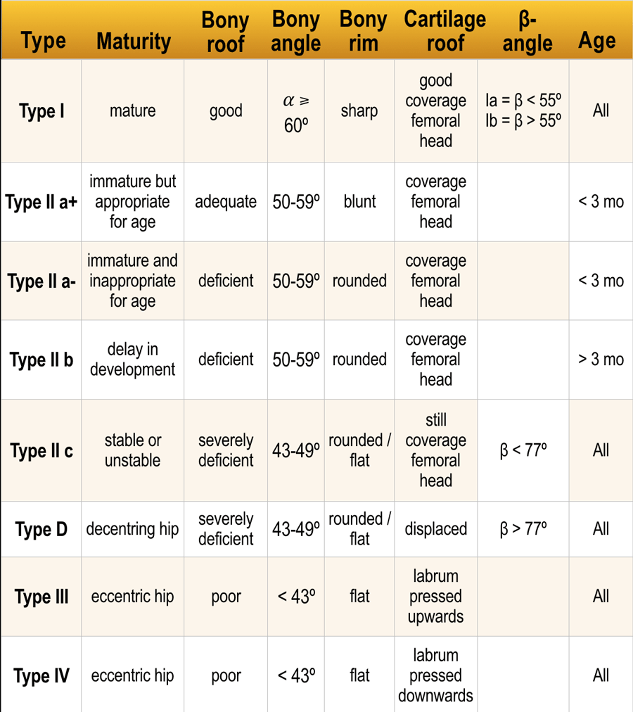
Graf's classification
- Type I:
Mature centred hip joint.
Well developed acetabular roof.
Angular or slightly blunt bony rim. - Type II:
Centred joint.
Deficiently developed acetabular roof Rounded bony rim - Type III:
Decentred joint.
Poorly developed acetabular roof. Flattened bony rim.
Click here for article on Developmental Dysplasia of the Hip.
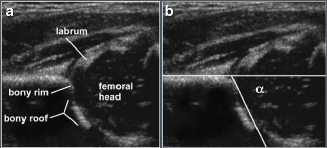
Normal ultrasonographic anatomy of the hip joint in the coronal plane (a).
Measurement of α angle (b)
Kidney
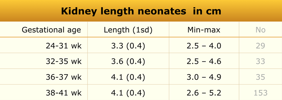 Adapted from reference 6
Preterm and Term babies
Material and methods
US study in 261 healthy newborn infants.
Craniocaudal dimension of the kidneys was determined with ultrasonography.
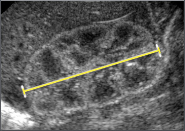
Ultrasonographic measurement of the length of a neonatal kidney.
Note the increased echogenicity of the renal parenchyma compared to liver parenchyma.
This is normal at this age.
 Adapted from reference 16
Children
Materials and method
Two hundred and three patients were included in this ultrasonography study.
Patients were excluded if they had a history of malignancy, use of steroids, upper urinary tract abnormality, VUR greater than grade I, urological surgery or if sonography of the kidney was regarded as abnormal.
On average the left kidney was 1.9 mm larger than the right kidney.
Causes of enlarged kidneys:
- Duplicated collecting system
- Nephritis (Infectious and non-infectious)
- Leukemia and Lymphoma
- ARPKCD
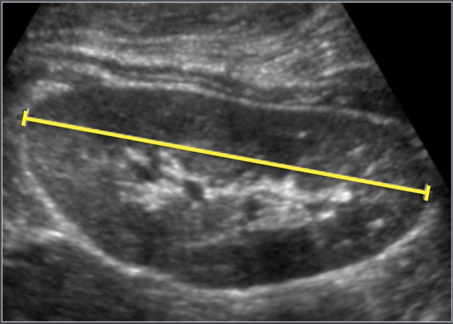
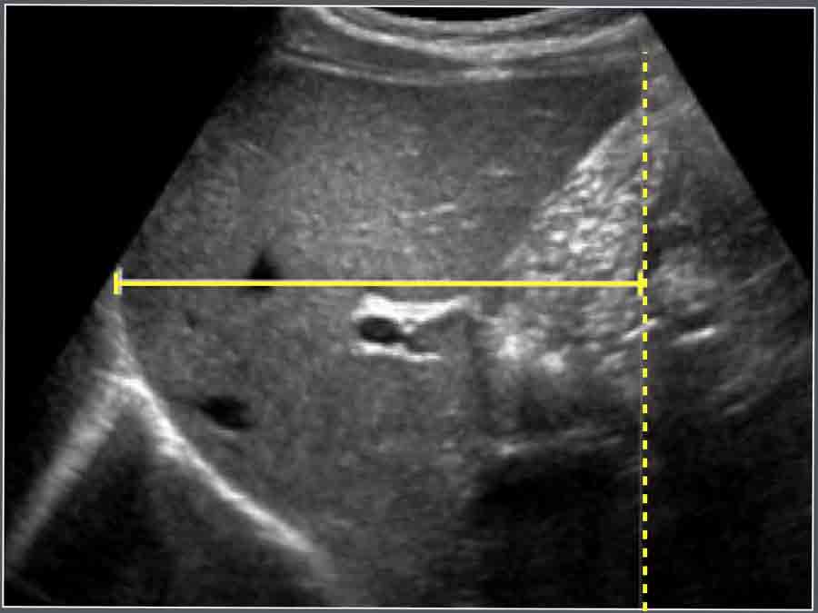
Ultrasonographic measurement of the length of a kidney.
 Adapted from reference 17
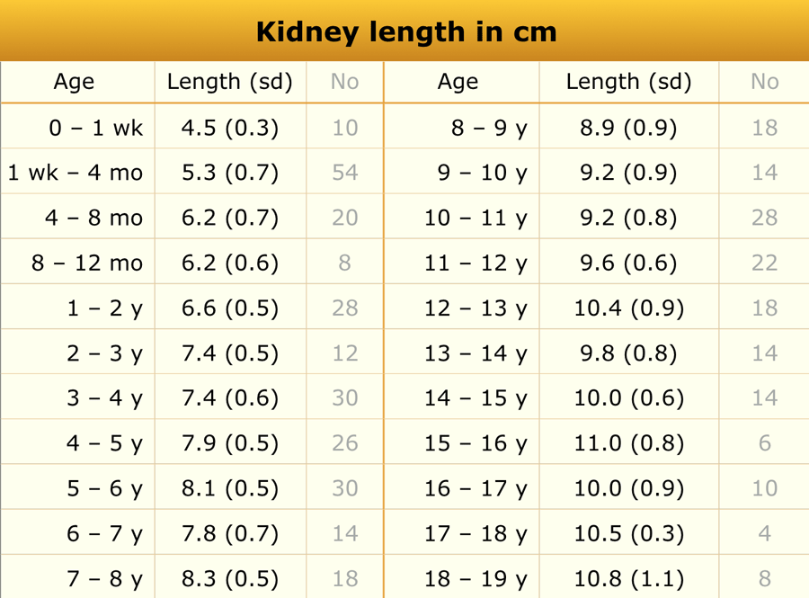
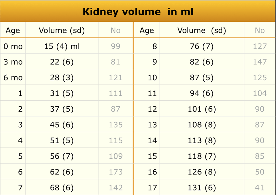
Kidney volume
Material and method
A total of 3376 children were recruited in this ultrasonographic study.
Kidney volume was calculated using the ellipsoid formula as Length x Width x Depth x 0.523.
In this study, the total renal volume was obtained by adding together both kidney volumes but without mentioning the separate values for the left and right kidney.
The values in the table were obtained by dividing the total renal volume by two.
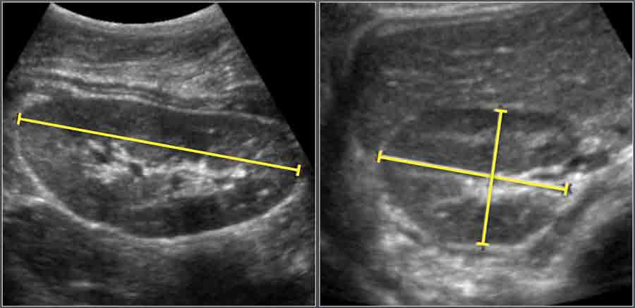
Ultrasonographic measurement of the length, width an depth of a kidney.
Kidney volume is calculated using the ellipsoid formula as Length x Width x Depth x 0.523.
 Adapted from reference 18
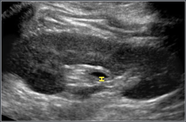
Thickness of the wall of the collecting system
Material and Methods
Ultrasonographic study of 48 renal collecting systems in 24 healthy children (age range 3 days to 12.6 years).
The collecting system could be identified in all kidneys and its wall thickness varied between 0 (not visible) and 0.8 mm.
Thickening of the wall ≥ 1mm is be considered as abnormal.
Causes of wall thickening:
- urinary tract infection
- intermittent dilatation (e.g. vesicoureteric reflux)
- dilatation in the recent past.
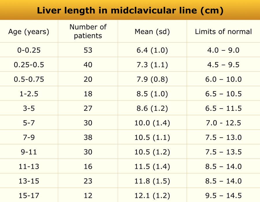
Liver
Craniocaudal dimension of the liver on the midclavicular line was measured with ultrasonography (figure).
Causes of hepatomegaly
- Leukemia
- Storage diseases
- (neonatal) Hepatitis
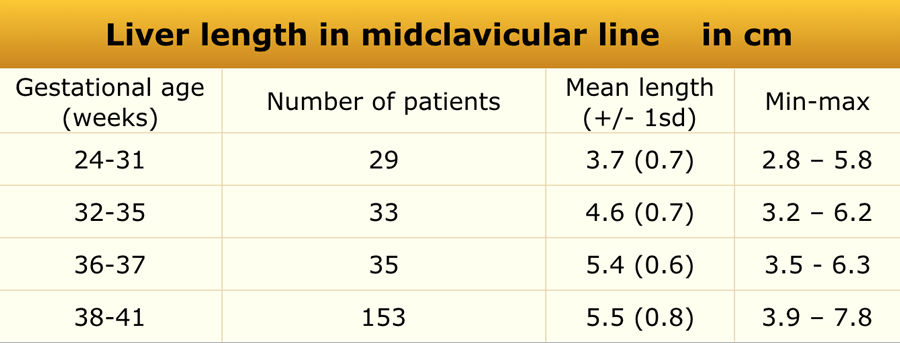 Adapted from reference 6
Newborns
Material and methods
US study in 261 healthy newborn infants. Craniocaudal dimension of the liver on the midclavicular line was determined with ultrasonography.
 Adapted from reference 7
Children
Material and methods
US study in 307 healthy children.
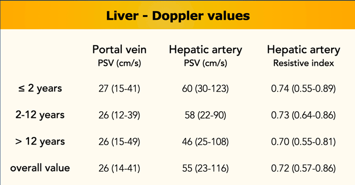
Doppler values
Materials and Method
One-hundred ultrasound examinations performed in 100 healthy children aged 0–17.9 years (median 7.8 years, interquartile range 1.1–14.1 years) were included.
Reference values for the hepatic hilum portal vein peak systolic velocity, hepatic artery peak systolic velocity, and hepatic artery resistive index in children were established (reference).
Portal vein peak systolic velocity is not age-dependent, whereas hepatic artery peak systolic velocity and hepatic artery resistive index decrease when children get older.
Mesenteric lymph nodes
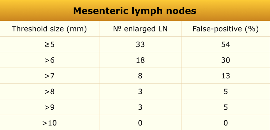 Adapted from reference 15
Materials and method
In this retrospective study in 61 children (36 boys and 25 girls, mean age 10.7 years, range 1.1-17.3 years) who underwent non-contrast abdominal CT examination for evaluation of suspected or known renal stones abdominal lymph node size was evaluated.
It is assumed that these CT findings can be extrapolated to ultrasonography.
Enlarged mesenteric lymph nodes (short axis > 5 mm) were found in 33 (54%) of the 61 children.
The majority of the enlarged mesenteric lymph nodes were found in the right lower quadrant (88%).
Based on their findings the authors state that: using a short-axis diameter of >8 mm might be a more appropriate definition for mesenteric lymphadenopathy in children.
False-positive rate for enlarged mesenteric lymph nodes with varying lymph node threshold size is seen in the table.
Pathologically enlarged lymph nodes:
- Intestinal lymphoma
- Lymphogenic metastasis
- Specific enteritis (e.g. TBC)
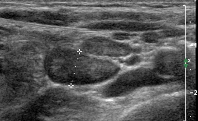
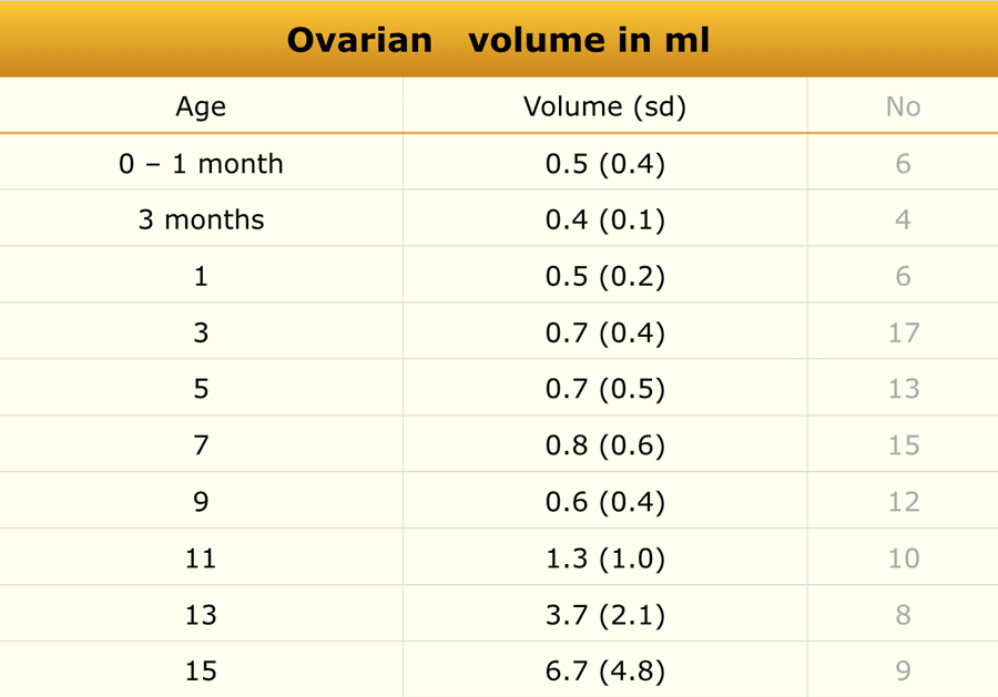
Ovary
 Adapted from reference 20
Material and Method
Ultrasonographic measurement of uterine and ovarian volume was performed in 178 healthy girls.
Causes of enlarged ovaries:
- Precocious puberty
- Ovarian torsion
- Polycystic ovarian disease
- Teratoma/dermoid
Ovarian volume is calculated using the formula:
- Length x Width x Height x 0.523.
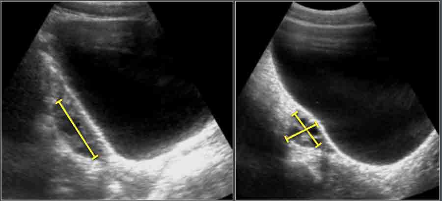
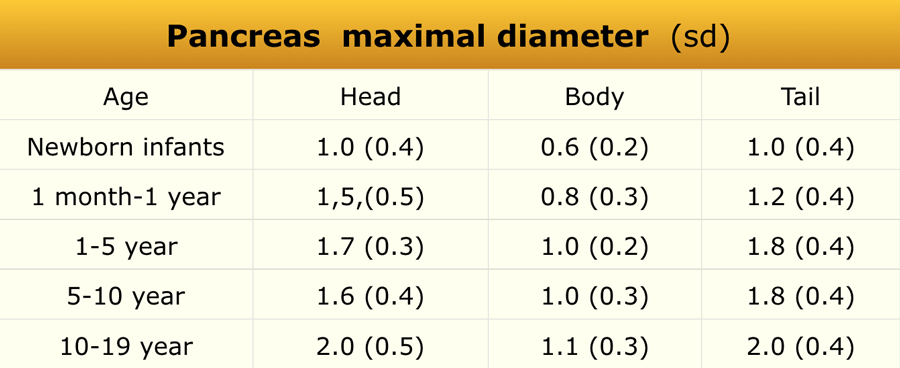
Pancreas
 Adapted from reference 12
Materials and method
Two hundred and seventy-three patients (differentiation in sex not mentioned) were included in this retrospective ultrasonography study.
The maximum anteroposterior (AP) diameters of the head, body and tail of the pancreas were measured on transverse/oblique images.
Echogenicity was low in 27 (10%), isoechoic in 145 (53%) and high in 101 (37%).
The maximum anteroposterior (AP) diameters of the head, body and tail of the pancreas were measured on transverse/oblique images.
Causes of enlargement of the pancreas:
- Traumatic pancreatitis
- Viral pancreatitis
- Drug-induced pancreatitis
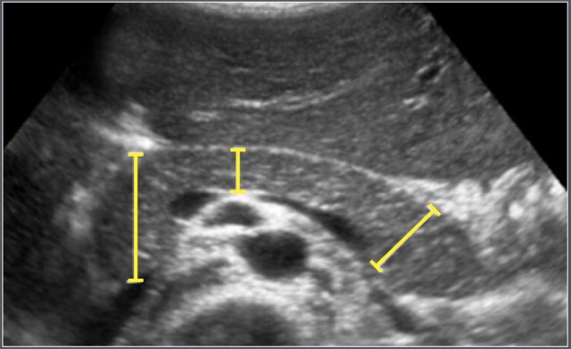
Portal vein
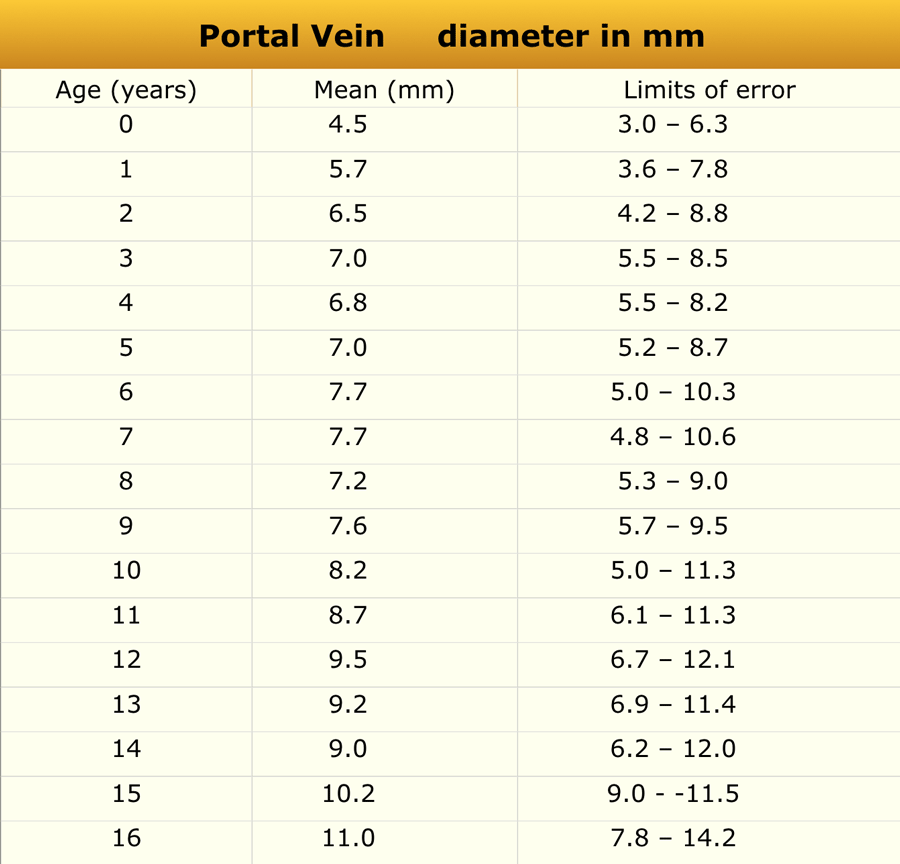 Adapted from reference 9
Materials and method
One hundred and fifty children aged 0-16 years, without clinical evidence of liver or intestinal disease, which were referred for abdominal ultrasound were included in the study.
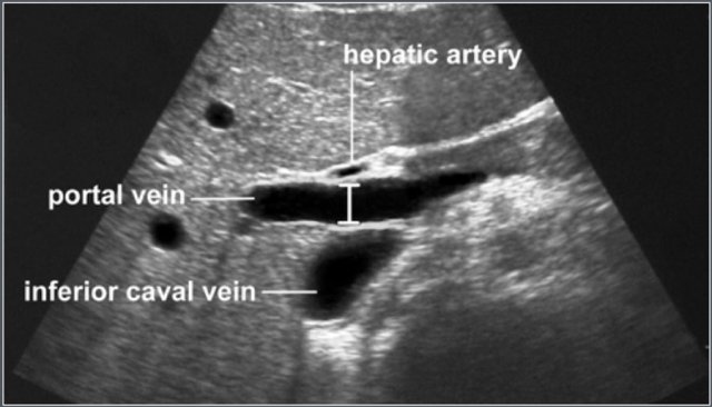 Measurement of portal vein diameter
The portal vein is visualized in the longitudinal axis from the splenomesenteric junction to the liver hilum.
The greatest anteroposterior diameter is measured at the site where the hepatic artery crosses the portal vein.
Spleen
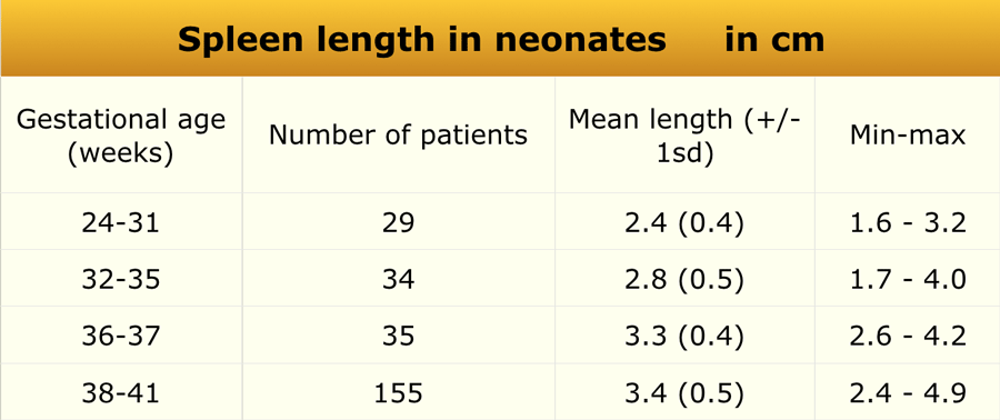 Adapted from reference 6
Preterm and term babies
Material and methods
US study in 261 healthy newborn infants. Craniocaudal dimension of the spleen was determined with ultrasonography.
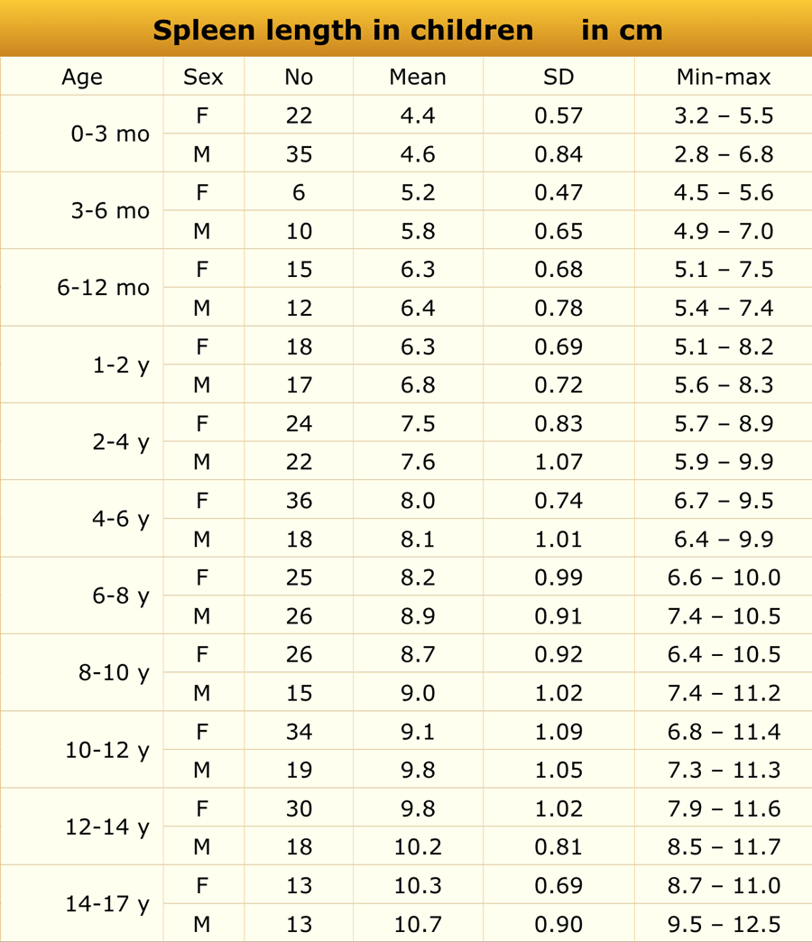 Adapted from reference 11
Children
Materials and method
These ultrasonography studies comprised of 512 healthy children - 238 boys and 274 girls - with ages ranging from 1 day (full-term neonate) to 17 years and 96 premature infants with gestational ages from 25-35 weeks.
None of the children had a problem that could affect spleen size.
Ultrasonography was performed using standard probes matched for age.
Causes of splenomegaly:
- Portal hypertension
- Leukemia
- Systemic infections (e.g. EBV or CMV)
- Hematologic disease (e.g. spherocystosis or thalassemia)
- Storage diseases
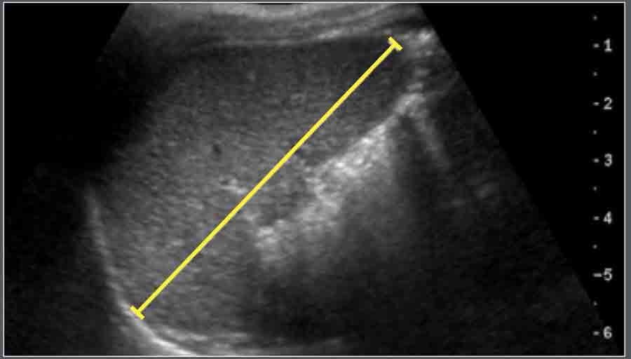
The measurement of spleen length is the optically maximal distance -ideally at the hilum - on the longitudinal coronal view between the most superomedial and the most inferolateral points (figure).
Subarachnoid space
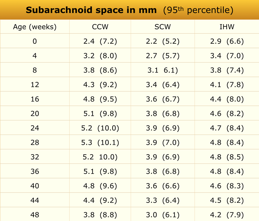 Adapted from reference 5
The subarachnoid space was assessed using ultrasonography in 278 full-term healthy Chinese infants. Measurements were taken in the coronal plane at the level of the foramen of Monro (figures)
The mean values in the table were calculated from the equations given in the article, the 95% confidence levels were derived from the graphs in the article.
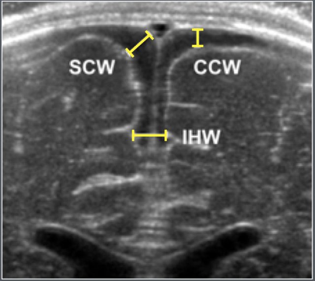
Ultrasonographic coronal representation of the subarchnoid space at the level of the foramen of Monro.
- SCW = Sino Cortical Width
- CCW = Cranio Cortical Width
- IHW = Inter Hemispherical Width.
Causes of enlargement of the subarachnoid spaces:
- BESSI (benign enlargement of subarachnoid space of infancy)
- Brain atrophy
- Dural sinus thrombosis
Testicle
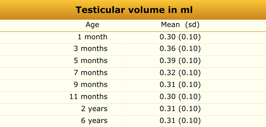 Adapted from reference 19
Materials and method
A total of 344 boys from different ethnic backgrounds were studied.
There were no differences between ethnic groups or between right and left testicle.
Causes of enlargement of the testis:
- Leukemia
- Precocious puberty
- Testicular torsion
- Epididymo-orchitis
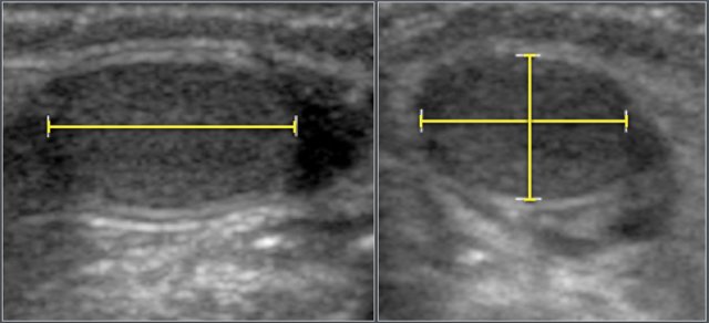
Testicular volume was calculated using the formula:
- Length x Width x Height x 0.523.
Thymus
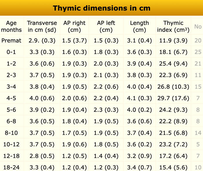 Adapted from reference 22
Materials and method
Mediastinal ultrasonography was performed in 151 infants (79 boys and 72 girls).
All children were healthy and had no stress factors affecting their thymic size.
Causes of enlarged thymus:
- Rebound thymus hyperplasia
- T-cell lymphoma of leukemia
- Thymoma
- Langerhans cell histiocytosis
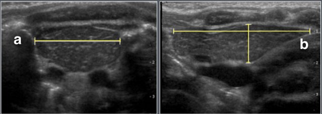
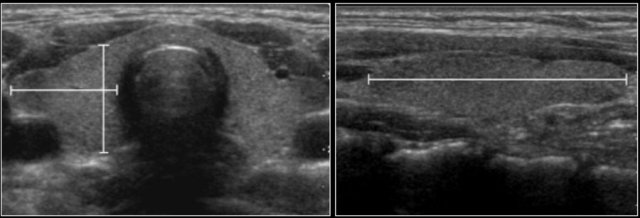
The maximum transverse diameter, right lobe anteroposterior, left lobe anteroposterior.
Perpendicular to the transverse plane the longest craniocaudal dimension (length) is assessed.
The thymic index was calculated by multiplying the transverse diameter (a) by the largest sagittal area (b).
Thyroid
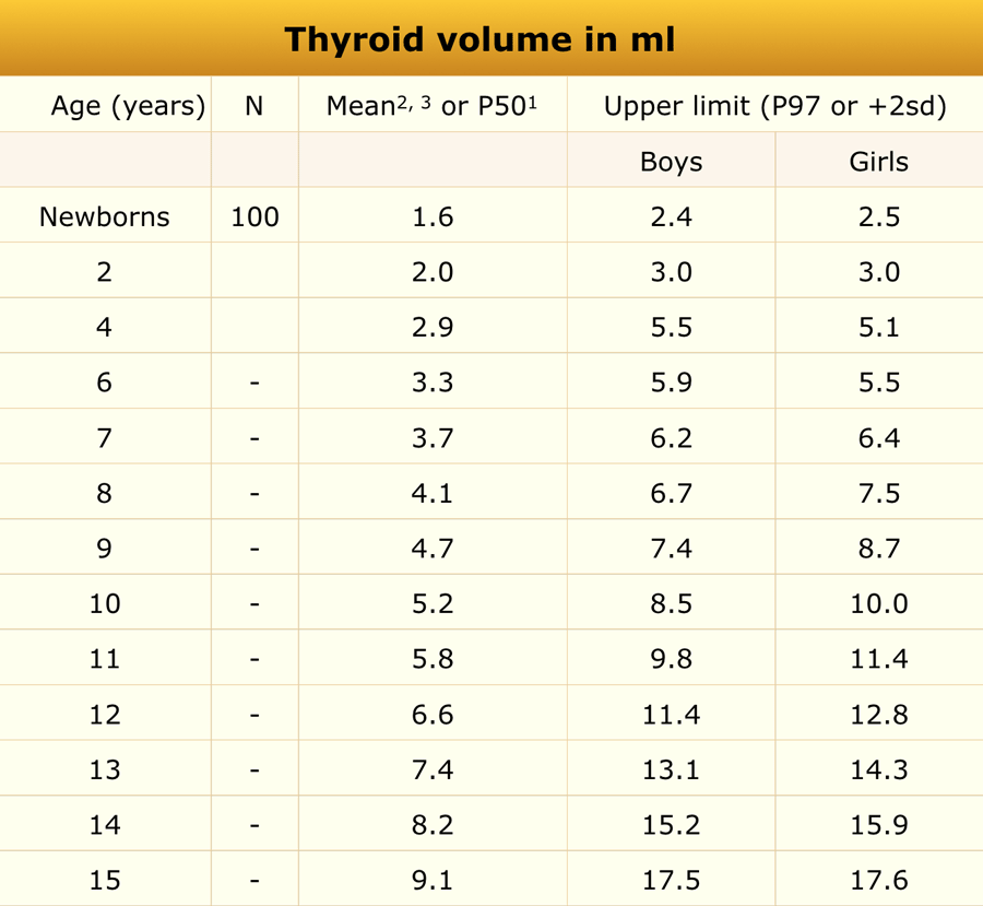 Adapted from reference 1-3
Material and methods
US study in 100 English newborn infants in the first week of life, a subset of iodine sufficient European children from a study of 5709 children, aged 6-15 years 1 and a subset of German children from a study of 252 children aged 2-4 years 2 [1-3].
The thyroid volume was the sum of the volumes of both lobes.
The volume of the isthmus was not included.
Causes of enlargement of thyroid gland:
- Thyroiditis (Hashimoto, Graves)
- Multinodular goiter
The volume of a thyroid lobe is calculated by the formula of an ellipsoid (length x width x height x 0,52).
Uterus
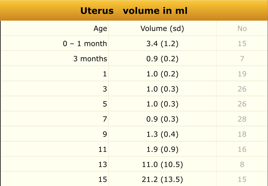 Adapted from reference 20
Material and Method
Ultrasonographic measurement of uterine and ovarian volume was performed in 178 healthy girls.
Causes of enlargement of uterus:
- Precocious puberty
- Hydro(hemato)metrocolpos
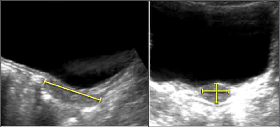
Uterine volume was calculated using the formula:
- Length x Width x Height x 0.523.
Ventricles
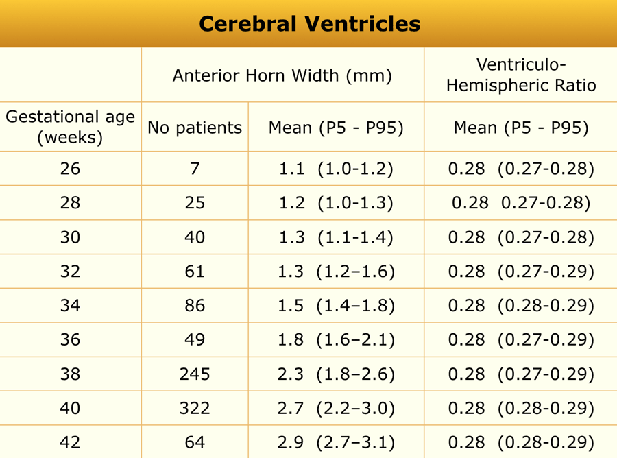 Adapted from reference 4
Adapted from an ultrasonographic study of 1483 neonates, gestational age range 25-42 weeks.
The neonates were examinded at day 3.
All neonates with perinatal asphyxia, infection of the central nervous system, intracranial hemorrhages of craniospinal malformation were excluded.
Causes of ventriculomegaly:
- Congenital (e.g. holoprosencephaly)
- Obstructive hydrocephalus
- Communicating hydrocephalus
- Atrophy
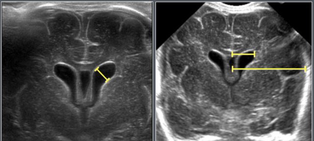
The anterior horn width and the ventriculo-hemspheric ratio is measured on the coronal view at the level of the foramen of Monro.