Temporal Bone Anatomy 2.0
Anatomy and related pathology
Erik Beek and Robin Smithuis
Radiology department of the University Medical Centre of Utrecht and the Alrijne Hospital in Leiderdorp, the Netherlands
Publicationdate
This is an updated version of the 2007 article.
In this review we present the normal axial and coronal anatomy of the temporal bone by scrolling through the images.
Some structures are discussed in more detail with emphasis on related pathology.
You will find more temporal bone pathology here.
Introduction
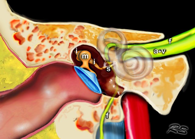
The middle ear consists of the tympanic cavity and the antrum.
The tympanic cavity is the major portion of the middle ear and contains the ossicles.
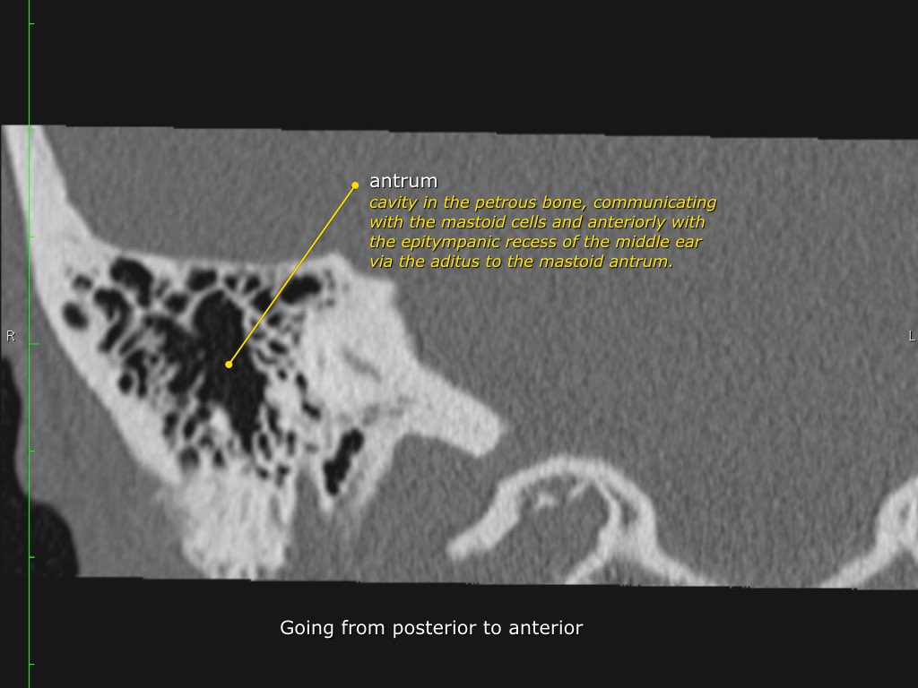
Via the aditus ad antrum the tympanic cavity is connected to the mastoid antrum, which is a large aircell superiorly and posteriorly to the tympanic cavity, and communicates with the mastoid air cells.
The tympanic membrane, the malleus, incus and stapes transfer soundwaves to the stapes footplate, which is attached to the base of the cochlea in the oval window.
Axial anatomy of petrous bone
Click on the image to enlarge.
Scroll through the images.
Coronal anatomy of petrous bone
Click on the image to enlarge.
Scroll through the images.
Tympanic cavity
The tympanic cavity is the major portion of the middle ear and contains the ossicles of the middle ear.
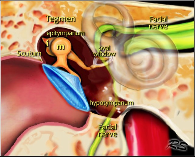
Medial wall
contains the oval and round window and the prominence of the tympanic segment of the facial nerve.
Lateral wall
is mainly formed by the tympanic membrane. The scutum is the bony prominence at the upper part or pars flaccidum of the tympanic membrane.
Roof
is called the tegmen and separates the upper part of the tympanic cavity or epitympanum from the middle cranial fossa.
Posterior wall
forms the entrance to the mastoid and is called the aditus ad antrum.
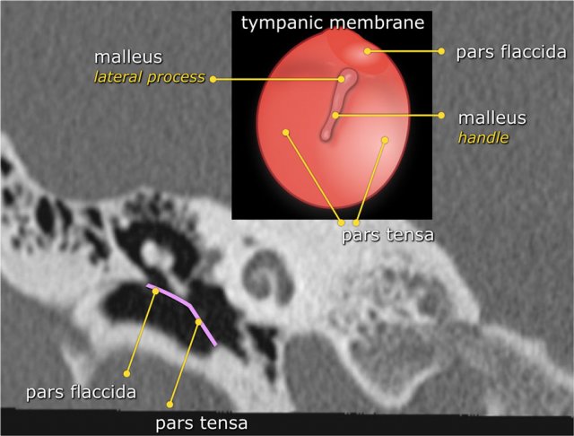
Tympanic membrane
The tympanic membrane or eardrum is a cone-shaped membrane that separates the external ear from the middle ear.
The pars flaccida is the upper fragile part that is associated with eustachian tube dysfunction and cholesteatoma.
The pars tensa is larger and more rubust and associated with perforations.
The handle or manubrium of the malleus is connected to the central part of the tympanic membrane, which is called the umbo.
 Perforation of the tympanic membrane.
Here a patient with a cholesteatoma.
There is a soft tissue mass in the epitympanum.
Notice the perforation of the tympanic membrane (yellow arrow) and the erosion of the lateral semicircular canal (red arrow).
The scutum is blunted.
 Erosion of scutum in a patient with a cholesteatoma. Also erosion of body of incus.
Scutum
This is usually the first bony structure to erode as a result of a cholesteatoma, that is formed by medial retraction of the pars flaccida of the tympanic membrane into the epitympanum.
If the retraction continues it will result in ossicular destruction.
If the cholesteatoma passes posteriorly through the aditus ad antrum into the mastoid itself, erosion of the tegmen mastoideum, with exposure of the dura and erosion of the lateral semicircular canal with deafness and vertigo, may result.
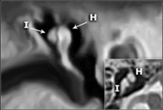
Ossicles
In many illustrations you will see the incus connecting medially to the malleus, but this is not correct.
On the coronal reconstruction on the left it is clearly demonstated that the incus is positioned posterolaterally to the malleolar head.
The long crus of the incus subsequently runs inferomedially to the stapes.
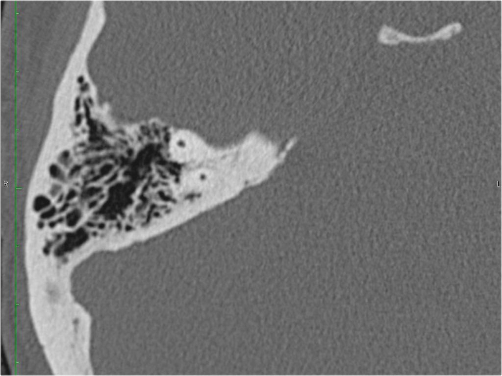
Antrum
The antrum is a large aircell superior and posterior to the tympanic cavity and connected to the tympanic cavity via the aditus ad antrum.
It is surrounded by smaller mastoid aircells.
Facial nerve
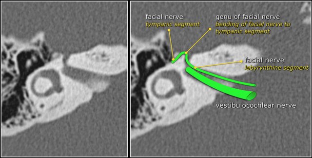
The labyrinthine segment of the facial nerve coming from the internal auditory canal angles sharply forward, nearly at right angles to the long axis of the petrous bone, to reach the geniculate ganglion.
At the ganglion the facial nerve makes a U-turn (first genu of the facial nerve) to run posteriorly as the tympanic segment along the medial wall of the epitympanum.
Oval and round window
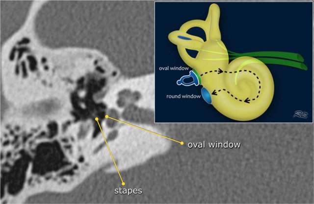
The base of the stapes rocks in and out against the membrane in the oval window.
The vibrations are transmitted from the oval window via the endolymph to the hair cells of the organ of Corti in the cochlea.
The round window dissipates the pressure generated by the fluid vibrations within the cochlea and thus serves as a release valve.