Coronary anatomy and anomalies
Robin Smithuis and Tineke Willems
Radiology department of the Rijnland Hospital Leiderdorp and the University Medical Centre Groningen, the Netherlands.
Publicationdate
In this article we describe the anatomy of the coronary arteries of the heart and some of the anomalies with illustrations and CT-images.
This article is an update of an article that appeared earlier in the Radiology Assistant.
Overview
 RCA, LAD and Cx in the anterior projection
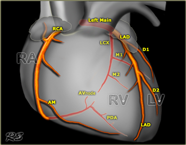
On the left an overview of the coronary arteries in the anterior projection.
- Left Main or left coronary artery (LCA)
-
Left anterior descending (LAD)
- diagonal branches (D1, D2)
- septal branches
-
Circumflex (Cx)
- Marginal branches (M1,M2)
-
Left anterior descending (LAD)
-
Right coronary artery
- Acute marginal branch (AM)
- AV node branch
- Posterior descending artery (PDA)
 RCA, LAD and Cx in the right anterior oblique projection
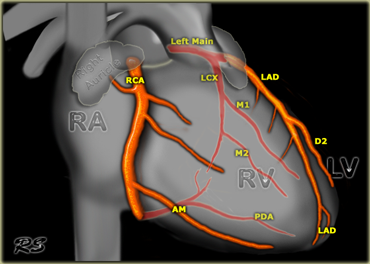
On the left an overview of the coronary arteries in the right anterior oblique projection.
- Left Main or left coronary artery (LCA)
-
Left anterior descending (LAD)
- diagonal branches (D1, D2)
- septal branches
-
Circumflex (Cx)
- Marginal branches (M1,M2)
-
Left anterior descending (LAD)
-
Right coronary artery
- Acute marginal branch (AM)
- AV node branch
- Posterior descending artery (PDA)
 RCA, LAD and Cx in the lateral projection
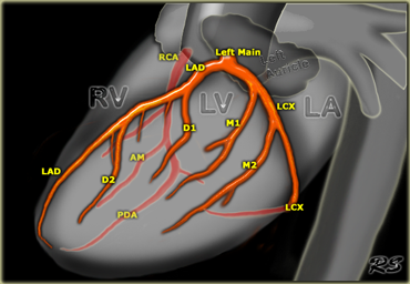
On the left an overview of the coronary arteries in the lateral projection.
- Left Main or left coronary artery (LCA)
-
Left anterior descending (LAD)
- diagonal branches (D1, D2)
- septal branches
-
Circumflex (Cx)
- Marginal branches (M1,M2)
-
Left anterior descending (LAD)
-
Right coronary artery
- Acute marginal branch (AM)
- AV node branch
- Posterior descending artery (PDA)
Read more about coronary anatomy inIntroduction to cardiothoracic imaging
Left Coronary Artery (LCA)
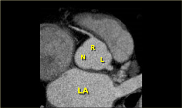 Left coronary (LC), right coronary (RC) and posterior non-coronary (NC) cusp
The left coronary artery (LCA) is also known as the left main.
The LCA arises from the left coronary cusp.
The aortic valve has three leaflets, each having a cusp or cup-like configuration.
These are known as the left coronary cusp (L), the right coronary cusp (R) and the posterior non-coronary cusp (N).
Just above the aortic valves there are anatomic dilations of the ascending aorta, also known as the sinus of Valsalva.
The left aortic sinus gives rise to the left coronary artery.
The right aortic sinus which lies anteriorly, gives rise to the right coronary artery.
The non-coronary sinus is postioned on the right side.
 LCA divides into LAD and Cx
The LCA divides almost immediately into the circumflex artery (Cx) and left anterior descending artery (LAD).
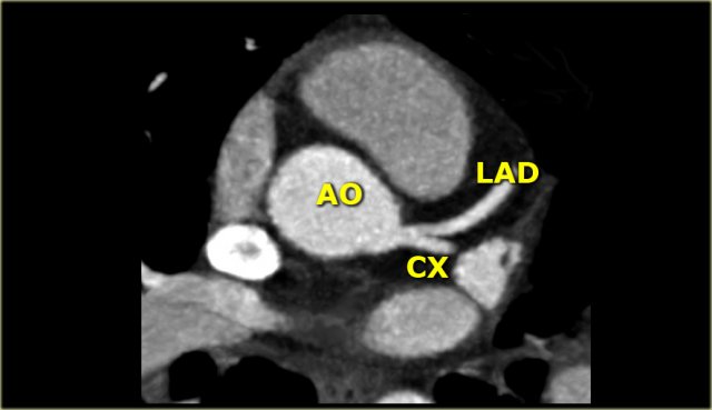
On the left an axial CT-image.
The LCA travels between the right ventricle outflow tract anteriorly and the left atrium posteriorly and divides into LAD and Cx.
On the image on the left we see the left main artery dividing into
- Cx with obtuse marginal branch (OM)
- LAD with diagonal branches (DB)
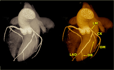
On volume rendered images the left atrial appendage needs to be removed to get a good look on the LCA.
In 15% of cases a third branch arises in between the LAD and the Cx, known as the ramus intermedius or intermediate branch.
This intermediate branche behaves as a diagonal branch of the Cx.
Left Anterior Descending (LAD)
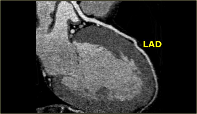 CT image of the LAD in RAO projection
The LAD travels in the anterior interventricular groove and continues up to the apex of the heart.
The LAD supplies the anterior part of the septum with septal branches and the anterior wall of the left ventricle with diagonal branches.
The LAD supplies most of the left ventricle and also the AV-bundle.
Mnemonic: Diagonal branches arise from the LAD.
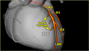
The diagonal branches come off the LAD and run laterally to supply the antero-lateral wall of the left ventricle.
The first diagonal branch serves as the boundary between the proximal and mid portion of the LAD (2).
There can be one or more diagonal branches: D1, D2 , etc.
Circumflex (Cx)
Circumflex and LAD seen in Lateral projection
The Cx lies in the left AV groove between the left atrium and left ventricle and supplies the vessels of the lateral wall of the left ventricle.
These vessels are known as obtuse marginals (M1, M2...), because they supply the lateral margin of the left ventricle and branch off with an obtuse angle.
In most cases the Cx ends as an obtuse marginal branch, but 10% of patients have a left dominant circulation in which the Cx also supplies the posterior descending artery (PDA).
Mnemonic: Marginal branches arise from the Cx and supply the lateral Margin of the left ventricle.
Right Coronary Artery (RCA)
RCA, LAD and LCx in Anterior projection
The right coronary artery arises from the anterior sinus of Valsalva and courses through the right atrioventricular (AV) groove between the right artium and right ventricle to the inferior part of the septum.
In 50-60% the first branch of the RCA is the small conus branch, that supplies the right ventricle outflow tract.
In 20-30% the conus branch arises directly from the aorta.
In 60% a sinus node artery arises as second branch of the RCA, that runs posteriorly to the SA-node (in 40% it originates from the Cx).
The next branches are some diagonals that run anteriorly to supply the anterior wall of the right ventricle.
The large acute marginal branch (AM) comes off with an acute angle and runs along the margin of the right ventricle above the diaphragm.
The RCA continues in the AV groove posteriorly and gives off a branch to the AV node.
In 65% of cases the posterior descending artery (PDA) is a branch of the RCA (right dominant circulation).
The PDA supplies the inferior wall of the left ventricle and inferior part of the septum.
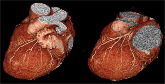
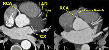 LEFT: RCA comes off the right sinus of ValsalvaRIGHT: Conus artery comes off directly from the aorta
On the image on the far left we see the most common situation, in which the RCA comes off the right cusp and will provide the conus branch at a lower level (not shown).
On the image next to it, we see a conus branch, that comes off directly from the aorta.
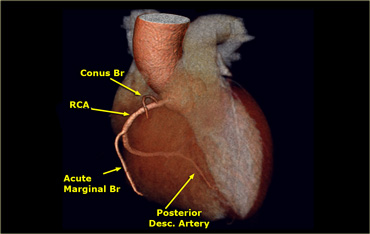
The large acute marginal branch (AM) supplies the lateral wall of the right ventricle.
In this case there is a right dominant circulation, because the posterior descending artery (PDA) comes off the RCA.
Coronary Anomalies
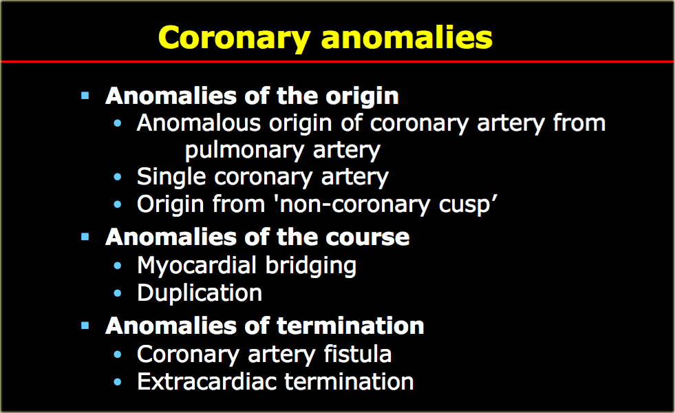
Coronary anomalies are uncommon with a prevalence of 1%.
Early detection and evaluation of coronary artery anomalies is essential because of their potential association with myocardial ischemia and sudden death (3).
With the increased use of cardiac-CT, we will see these anomalies more frequently.
Coronary anomalies can be differentiated into anomalies of the origin, the course and termination (Table).

The illustration in the left upper corner is the most common and clinically significant anomaly.
There is an anomalous origin of the LCA from the right sinus of Valsalva and the LCA courses between the aorta and pulmonary artery.
This interarterial course can lead to compression of the LCA (yellow arrows) resulting in myocardial ischemia.
The other anomalies in the figure on the left are not hemodynamically significant.
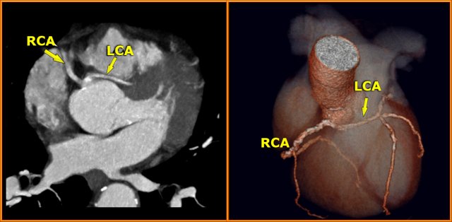
Interarterial LCA
On the left images of a patient with an anomalous origin of the LCA from the right sinus of Valsalva and coursing between the aorta and pulmonary artery.
Sudden death is frequently observed in these patients.
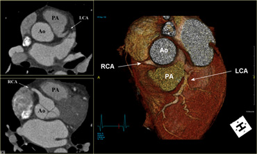
ALCAPA
On the left images of a patient with an anomalous origin of the LCA from the pulmonary artery, also known as ALCAPA.
ALCAPA results in the left ventricular myocardium being perfused by relatively desaturated blood under low pressure, leading to myocardial ischemia.
ALCAPA is a rare, congenital cardiac anomaly accounting for approximately 0.25-0.5% of all congenital heart diseases.
Approximately 85% of patients present with clinical symptoms of CHF within the first 1-2 months of life.
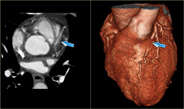
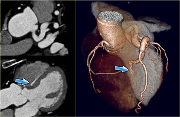
Myocardial bridging
Myocardial bridging is most commonly observed of the LAD (figure).
The depth of the vessel under the myocardium is more important that the lenght of the myocardial bridging.
There is debate, whether some of these myocardial bridges are hemodynamically significant.
 Left to right shunt: septal branch of LAD teminates in right ventricle
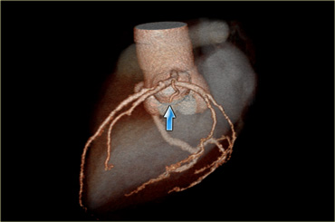
Fistula
On the image on the left we see a large LAD giving rise to a large septal branch that terminates in the right ventricle (blue arrow).