Aortic Aneurysm Rupture
CT signs of pending aortic aneurysm rupture
Jay P. Heiken, M.D.
Mallinckrodt Institute of Radiology of the Washington University School of Medicine, St. Louis, Missouri
Publicationdate
This article is based on a presentation given by Jay Heiken and adapted for the Radiology Assistant by Robin Smithuis.
Jay Heiken is professor of radiology with special interest in abdominal imaging and co-author of the well known book 'Computed Body Tomography With Mri Correlation'.
The classical findings in aortic aneurysm rupture are well known.
In this article we will present the more subtle findings of contained leak and pending rupture of aortic aneurysm.
Primary signs of Aortic Aneurysm rupture

Aortic aneurysm rupture is the most important diagnosis you want to be able to exclude in patients with acute abdominal pain especially when they present with back or flank pain.
The primary signs of AAA rupture are periaortic stranding, retroperitoneal hematoma and extravasation of iv. contrast.
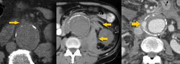 LEFT: Subtle periaortic stranding, MIDDLE: Hemorrhage into posterior pararenal and perirenal compartment, RIGHT: Extravasation of iv. contrast
On the left we see three patients with aortic aneurysm rupture.
In the image on the far left we only see a little bit of peripheral soft tissue density adjacent to the aneurysm and indeed this is a sign that this patient is at risk for frank rupture.
The other two cases show retroperitoneal hematoma and contrast leakage outside the aorta, which makes it easier to diagnose.

On the left a classical case in a patient with an aneurysm of the abdominal aorta and a large hyperdense retroperitoneal hematoma due to rupture.
The majority of these cases show posterior periaortic hemorrhage and in cases of massive hemorrhage, the posterior pararenal and perirenal compartments are the most frequently involved sites.
Signs of Pending Aneurysm Rupture

The CT features of contained leak or pending rupture of an aortic aneurysm may be subtle and easily overlooked.
We have to look for the high-attenuating crescent sign, focal discontinuity of intimal calcification or tangential calcium or the draped aorta sign (table on the left).
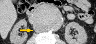
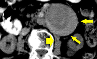 High-attenuating crescent sign in a patient with subtle evidence of leak adjacent to the right psoas muscle (broad arrow).
High-attenuating crescent
The high attenuating crescent represents an acute hematoma within either the mural thrombus or the aneurysmal wall.
This sign is strongly associated with AAA rupture.
Sensitivity of the high-attenuating crescent sign as an indication of complicated aneurysm is 77%; specificity, 93%; and positive predictive value of 53%.
So even if there are no primary signs of rupture, we need to indicate to the referring physician, that this patient is at very high risk for aneurysm rupture within the next few days.
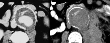
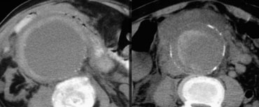 High-attenuating crescent sign
On the left two more cases of the high-attenuating crescent sign.
In the case on the right we can also identify a retroperitoneal hematoma, so in this case there is a frank AAA rupture.

Focal discontinuity of intimal calcification
Another sign of impending rupture or contained leakage is focal discontinuity of intimal calcification.
In most of these cases we can also identify the tangential calcium sign.
In these cases it looks as if the calcium is pointing out away from the expected circumference of the aneurysm.
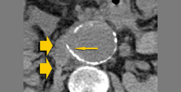 Tangential calcium sign (small arrow) and hemorrhage (broad arrow)
Tangential calcium sign
On the left we see another example of the tangential calcium sign.
The intimal calcification points away from the aneurysm and there is retroperitoneal leakage.
 LEFT: draped aorta sign.RIGHT: two weeks later there is a rupture
Draped Aorta
On the left a patient who presented with backpain.
The image on the far left shows a bulge of the aorta.
This either represents a focal weakening of the aortic wall or a localized leak.
This patient unfortunately was reported as having no leak and discharged from the emergency departement.
Two weeks later there was a frank rupture.
A positive aortic drape sign is considered to be present when the following features are seen:
- area in which the posterior aortic wall is unidentifiable as a distinct line.
- the posterior aorta follows the contour of the spine on one or both sides.
 Draped aorta sign. Patient died 3 hours later.
On the left another patient who presented with backpain.
There was no evidence of aneurysm leakage, but we see a draped aorta.
The posterior contour of the aorta follows the contour of the spine as if the aorta is draped over the vertebral body.
There is no imaging follow up in this patient, but three hours after this image was taken, this patient exsanguinated from a ruptured AAA.
The leakage was probably where the bulge was (arrow).